Locations:

Symptoms complement one another
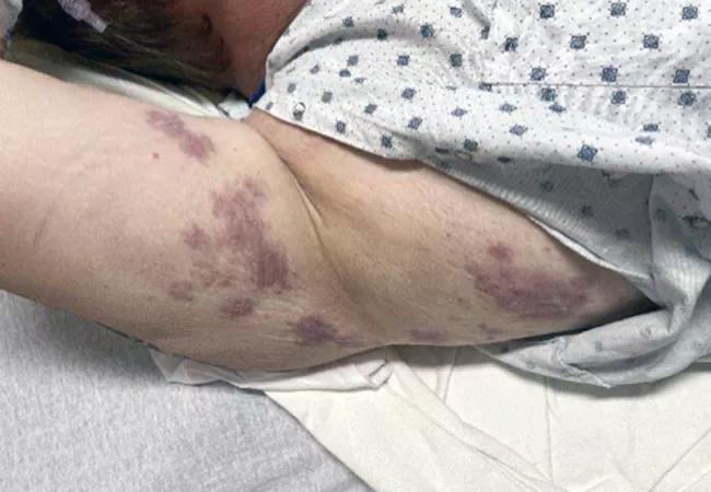
Image content: This image is available to view online.
View image online (https://assets.clevelandclinic.org/transform/6a35a0ae-8757-4047-9bc3-1edd14388984/23-RHE-4333860-CQD-650x450-1_jpg)
Skin lesions
Above: The patient’s raised lesions left hyperpigmented areas that looked like bruising.
Advertisement
Cleveland Clinic is a non-profit academic medical center. Advertising on our site helps support our mission. We do not endorse non-Cleveland Clinic products or services. Policy
by Komal Ejaz, MD, and Adam Brown, MD
We present the case of a man in his 40s who came to Cleveland Clinic for evaluation of inflammatory arthritis, rash, facial swelling, abdominal pain and weight loss. The patient was in his usual state of health until October 2022, when he developed inflammatory pain involving small joints of the hands and feet. He was found to have rheumatoid factor, an anti-cyclic citrullinated peptide positivity, along with likely rheumatoid nodules on his elbows. He was diagnosed with rheumatoid arthritis and started on methotrexate, but couldn’t tolerate it and was transitioned to leflunomide.
Within weeks, the patient developed further complications of multiple raised skin lesions and erythematous plaques with central clearing, which was present for multiple days, and left hyperpigmented, ecchymotic-appearing lesions. Along with the skin rash, he developed episodes of facial and tongue swelling and severe abdominal pain.
He was evaluated at an outside hospital, where he was diagnosed with angioedema. Imaging of his abdomen at the time revealed mural thickening through the ileum, duodenum and ascending colon. He underwent esophagogastroduodenoscopy and colonoscopy with biopsy without a clear diagnosis; angioedema of the gut was thought to be the most likely process. He was given glucocorticoids and fresh frozen plasma with some improvement in symptoms. No cause of the angioedema was found and no diagnosis for the rash was made at this time.
The patient was discharged with a prednisone taper, but his rash and abdominal pain persisted. Loss of appetite had led to a 40-pound weight loss within a span of three months. This prompted him to be
admitted to Cleveland Clinic, where he was evaluated by multiple specialties, including internal medicine, rheumatology, gastroenterology, dermatology and allergy/immunology.
Advertisement
During the patient’s second hospitalization, he continued to have joint pain, mostly in his hands, wrists and ankles. He did not have any fresh skin lesions; only the hyperpigmented lesions remained. Dermatology clinicians made a major contribution when they speculated that the lesions were most likely urticarial, despite not migrating and lasting for more than 24 hours. A biopsy revealed leukocytoclastic vasculitis. Considering the appearance and characteristics of the lesions and the
histological findings, a diagnosis of urticarial vasculitis was made. Additional testing revealed low C3 and C4, further classifying the diagnosis as hypocomplementemic urticarial vasculitis, potentially secondary to seropositive rheumatoid arthritis.
The patient had normal renal function and normal urinalysis. He was treated with high-dose glucocorticoids and dapsone, leading to rapid resolution of his symptoms, including abdominal pain.
We all learn in medical school that if you encounter a patient with an urticarial rash and examine them
sometime later, the rash will look different because classic urticaria moves from one place to another on the body. Urticarial vasculitis, however, is unique. It remains fixed in place for more than 24 hours, and as it resolves it leaves an area of hyperpigmentation. A biopsy of the rash, even as it’s resolving, can establish the diagnosis of urticarial vasculitis.1
Urticarial vasculitis can occur without an underlying condition, but, importantly, urticarial vasculitis is also associated with underlying systemic autoimmune conditions, such as systemic lupus erythematosus and, rarely, rheumatoid arthritis, as occurred in our patient.
Advertisement
Once a diagnosis of urticarial vasculitis is made, it can be divided into normocomplementemic urticarial vasculitis (NUV) or hypocomplementemic urticarial vasculitis (HUV) based on serum C3 and C4 levels. The distinction is important, as HUV tends to have more systemic involvement compared to NUV. Like in the patient described, HUV can be associated with angioedema and arthritis, but other systemic manifestations also can occur, such as glomerulonephritis and uveitis. NUV, in contrast, tends to be confined to the skin.
Angioedema is a striking feature of HUV. It can be life-threatening and seems to have a relatively unique association with HUV, as it is not commonly seen in other systemic autoimmune processes. The mechanism of angioedema is unclear but is thought to be potentially related to the production of C5a, which could lead to mast cell degranulation. C5a is produced because of the presence of anti-C1q antibodies forming immune complexes with C1q, triggering the classical complement cascade and depleting serum C3 and C4 in the process. C5a is produced in many other conditions that activate complements that are not associated with angioedema, so this is likely not the sole explanation for angioedema in this patient population. Anti-C1q antibodies can be ordered when HUV is suspected as they are often present.
Standard treatment of HUV is not established, but the literature shows that many medications have been used with reasonable success. Initially, as with many of our cutaneous conditions, colchicine or dapsone can be attempted along with antihistamines to help with the angioedema. More aggressive therapy includes disease-modifying anti-rheumatic drugs (DMARDS) and potentially rituximab.2
Advertisement
Urticarial vasculitis is different than chronic spontaneous urticaria. Urticarial vasculitis is fixed, lasting for greater than 24 hours, and leaves an area of hyperpigmentation upon resolution.
References
Advertisement
Advertisement

Protein expression in synovial fluid indicates patients’ immune factors may be involved

Collaboration must cross borders and disciplines

Clinical complexity demands personalized care strategies

Higher than 5-year rates for prostate cancer and melanoma and about the same as breast cancer

Sinus tracts can occur years later and not near the incision site
Researchers hope it may one day help patients avoid explantation surgery

Evidence suggests empathy influences immune function and well-being

Going beyond simple referrals to streamline patient access