Multilevel cervical fusion restores function in an athletic 78-year-old
Over a period of several years, a 78-year-old woman experienced changes in her cervical spine that compromised her overall function and quality of life. Extremely fit and formerly accustomed to hiking and playing golf, she presented to Cleveland Clinic’s Center for Spine Health in 2020 with severe neck pain and an inability to hold her head upright for prolonged periods.
Advertisement
Cleveland Clinic is a non-profit academic medical center. Advertising on our site helps support our mission. We do not endorse non-Cleveland Clinic products or services. Policy
Based on the findings of a physical examination and measurement of spinal curvature, spine surgeon Thomas Mroz, MD, diagnosed the patient with degenerative cervical kyphosis.
Also known as “bent forward deformity,” cervical kyphosis is estimated to occur in 20% to 40% of adults aged 60 or older. Symptoms include neck pain, myelopathy, radiculopathy, issues with horizontal gaze and, in severe cases, problems with swallowing and breathing. With age, cervical lordosis can be lost, and genetics, trauma, and smoking also have been shown to contribute to development of the condition.
Although the prevalence of cervical kyphosis in older adults is increasing with the aging of the U.S. population, surgery for these patients is uncommon, except in extreme cases. Careful preoperative assessment is necessary, with static and dynamic cervical radiography, to measure the deformity and for procedure planning.
“My decision-making process is aimed at giving patients the most durable benefits and ensuring, with a high degree of certainty, that surgery will be making them functionally better,” says Dr. Mroz.
After evaluation of the patient and an in-depth discussion about her struggles with symptoms related to cervical kyphosis and expectations for an outcome, Dr. Mroz determined that she was an appropriate surgical candidate.
“Had this patient not been so active and mentally alert, I might not have performed surgery,” he explains. “In fact, she had been seen at another institution, where they declined to operate. But I believed it was possible to balance the benefits and risks and make a meaningful change in her quality of life.”
Advertisement
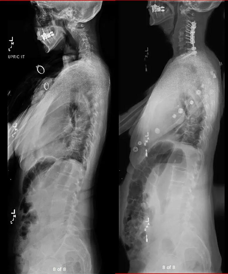
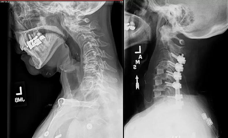
The patient underwent an anterior cervical discectomy fusion from C4 to T1 and posterior column osteotomies and fusion from C3 to T1. Figures 1 and 2 present pairs of preoperative and postoperative imaging studies showing the deformity correction. Dr. Mroz notes that the operation required “nuance to the technique, which enabled correction of the patient’s deformity. Had we not backed up the anterior procedure with additional posterior screws and rods, she may have gone on to develop a nonunion at one of the disk spaces, with loss of the correction.”

Image content: This image is available to view online.
View image online (https://assets.clevelandclinic.org/transform/39b842f3-c45b-4fb4-8ede-3076655d2dd7/22-NEU-3097423-CQD-Inset1-800x961-1_jpg)
Figure 1. Preoperative (left) and postoperative (right) full-length spine X-rays demonstrating correction of cervical kyphosis.

Image content: This image is available to view online.
View image online (https://assets.clevelandclinic.org/transform/38e6e75a-01a7-4abf-833a-34f3b5120023/22-NEU-3097423-CQD-Inset2-800x483-1_jpg)
Figure 2. Preoperative X-ray (left) and postoperative CT (right) demonstrating anterior and posterior correction of cervical kyphosis.
There is heterogeneity in surgical technique for adult cervical kyphosis from institution to institution, Dr. Mroz points out, because of a lack of standardized management guidelines. He notes that patients treated for such a deformity at Cleveland Clinic benefit from a multidisciplinary team approach that includes surgeons, medical spine and pain specialists, and other professionals who specialize in spine care.
More than two years out from surgery, the now 80-year-old patient was seen by Dr. Mroz for follow-up in June 2022. “She reported feeling great, having no neck pain or neurologic symptoms, and being back to playing competitive golf,” he says. “This was a really good outcome due, in part, to our ability to individualize her care and apply machine learning to data from this patient to assess her suitability for surgery.”
Advertisement
The technology to which Dr. Mroz alludes is the Center for Spine Health’s artificial intelligence-driven decision platform. With it, surgeons are able to analyze variables from the electronic medical record such as patient demographics, comorbidities, medications, laboratory test results and functional data. The result is an improved ability to predict the likelihood of success with a procedure.
The key takeaway from this case, Dr. Mroz says, is the need to empower patients with spine deformities to seek personalized approaches. “Many options are available for treatment,” he observes. “It’s important for clinicians to really listen to their patients, be aware of all the options that are available, and make referrals if necessary and appropriate.”
Advertisement
Advertisement

Aim is for use with clinician oversight to make screening safer and more efficient

Rapid innovation is shaping the deep brain stimulation landscape

Study shows short-term behavioral training can yield objective and subjective gains

How we’re efficiently educating patients and care partners about treatment goals, logistics, risks and benefits

An expert’s take on evolving challenges, treatments and responsibilities through early adulthood

Comorbidities and medical complexity underlie far more deaths than SUDEP does

Novel Cleveland Clinic project is fueled by a $1 million NIH grant

Tool helps patients understand when to ask for help