Locations:

A team of surgeons helped give a 9-year-old patient with congenital facial paralysis the ability to smile
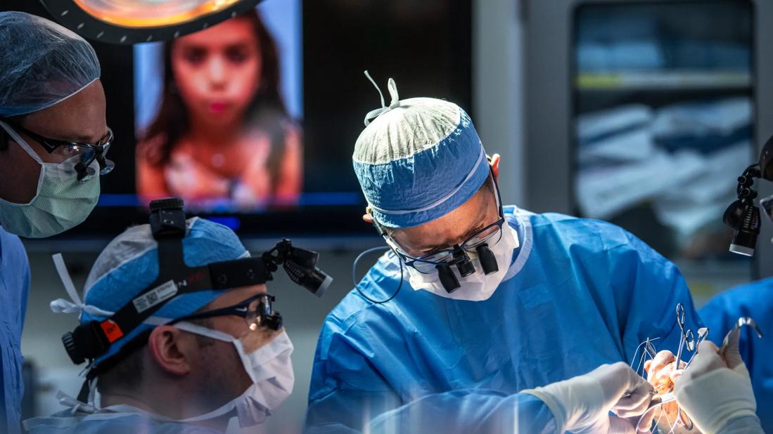
Image content: This image is available to view online.
View image online (https://assets.clevelandclinic.org/transform/69603611-5eca-4061-9dde-ecb8b5c86bfc/CCC_2191938_06-08-21_349_AMO)
Before and after images of patient with trivector trasoral trasfacial OTTT.
In 2019, a 7-year-old patient presented to Patrick Byrne, MD, MBA, the chairman of Cleveland Clinic’s Head & Neck Institute with congenital facial paralysis. She was unable to move the right side of her face, which impaired her ability to express emotion, smile or close her right eye. Two year later, she followed up in clinic with Dr. Byrne and also Dane Genther, MD, a facial plastic and reconstructive surgeon in Cleveland Clinic’s Head & Neck Institute, who specializes in the treatment of facial paralysis. Because of her lack of smile and weak lower eye lid, she was determined to be a good candidate for facial reanimation with the use of a gracilis free muscle transfer. This microvascular surgical procedure has become a gold standard for the management of longstanding facial paralysis. However, historically it has been performed with a single muscle belly, which is placed in a manner that allows elevation of the corner of the mouth. Dr. Byrne and others have in recent years developed novel innovations in an effort to more accurately recreate natural facial expression. This includes the use of multiple motor nerves, and multiple slips of muscle. Most recently, they have been developing “tri vector” flaps: in which not only multiple smile vectors are achieved, but also emotional movement around the eyes. To our knowledge, this is a unique approach worldwide, not only in adults, but now in children.
Advertisement
Cleveland Clinic is a non-profit academic medical center. Advertising on our site helps support our mission. We do not endorse non-Cleveland Clinic products or services. Policy
Dr. Byrne and Dr. Genther performed her facial reanimation procedure as a closely coordinated team. Dr. Byrne prepared the face and elevated tissue to get access to the nerves. He also elevated the skin and the fat all the way to the corner of the mouth to allow for placement of the flap. While Dr. Byrne focused on the patient’s face, Dr. Genther harvested the sural nerve from the patient’s lower leg for the cross-facial nerve graft and harvested the gracilis free flap from her thigh. The rest of the surgery was performed in collaboration to carefully position and secure the flap, using microsurgical techniques to reconstruct the blood vessels and nerves. Brandon Hopkins, MD, a Cleveland Clinic pediatric head and neck surgeon, consulted during the surgery.
The surgery took 12 hours to complete, and the patient responded well. She spent four days in the hospital for careful monitoring before being released home. Swelling in the face remained for a few weeks, which was expected. Although the patient does not have movement yet, which was also expected, Drs. Byrne and Genther anticipate that she should have movement and nerve signal by around four months after surgery based on their experience with previous patients who underwent tri-vector reanimation.
While the tri-vector gracilis technique has been employed and described in adult patients, to the team’s knowledge, this is the first instance of the tri-vector reanimation technique being used in a pediatric patient.
The gracilis free flap is the most complex procedure in facial reanimation, but it offers the greatest benefit to patients with severe facial paralysis. For patients who have no functioning facial muscles and/or nerves, the technique can restore movement by replacing the muscles and providing signal from alternative nerves.
Advertisement
“The gracilis microneurovascular free flap operation initially started out as just a single vector where a single slip of muscle would pull the lip up and out, but it didn’t do things like show the upper teeth, adequately elevate the upper lip or provide any movement around the eyes,” explains Dr. Genther. “It made your lip move like you were smiling, but it didn’t look natural, and it didn’t create a natural kind of Duchenne smile. To get more of a natural appearance, you need more vectors. Normally, there are six different muscles that move the lip up and out, and trying to achieve all of that with one vector just doesn’t result in as natural of an appearance.”
In both pediatric and adult patients, the surgery is optimally performed with a two-team approach. One surgeon, in this case, Dr. Byrne, prepares the face and provides access to all necessary structures, including nerves, blood vessels and locations where the flap will be secured. The incisions used to provide this access are carefully placed to allow them to fade and camouflage once they heal.
The other surgeon, Dr. Genther for this particular case, harvests the sural nerve for nerve grafting and the gracilis free flap, which is harvested with its associated nerves and blood vessels so blood flow and nerve signal can be restored once placed in the face.
As a team, the gracilis free flap is then prepared by separating the muscle fibers into different vectors to restore different aspects of the smile. Once prepared, the flap is inset to provide ideal facial movement. A microscope is then used to meticulously connect all nerves and blood vessels using suture thinner than a human hair.
Advertisement
Dr. Genther explains that each vector in this technique has a specific role. One vector is used to move the lip up and out, a shorter one provides additional elevation, and the third vector is around the eye to create the appearance of a genuine smile. Dr. Genther points to Dr. Byrne as one of the people who helped pioneer the tri-vector technique and getting more complex with the procedure. “In terms of pediatrics, as far as we know, this was the only tri vector flap in a child,” says Dr. Genther. “This technique has been around for about five years, and it’s been done in adults. But, in general, most people aren’t doing it; it’s just Dr. Byrne and maybe a few other people across the country.”

Image content: This image is available to view online.
View image online (https://assets.clevelandclinic.org/transform/27b3e2dd-7716-4d92-b315-635c9214aa6d/21-HNI-2202209-TriVector-transoralTansfacial-OTTT-CQD-hero_650x450-2-jpg)
Before and after surgery
The best candidates for this procedure are those with complete facial paralysis on one side. It is also an option for patients with incomplete paralysis but don’t have an adequate smile and don’t seem to have recoverable facial muscles. Dr. Genther notes that while all facial paralysis patients are potentially candidates for the dual- and tri-vector, a surgeon would not do a tri-vector if the patient had good eye movement. “The purpose of the third vector is to provide lower eyelid movement that is characteristic of a natural spontaneous smile,” says Dr. Genther. “With a Duchenne smile, your lower eyelids will wrinkle a bit. When your eyes don’t wrinkle, observers notice that movement is missing, and it can appear like you’re giving an inauthentic smile.”
Beyond improving a patient’s smile, Dr. Genther points out some additional advantages for the multi-vector reanimation. Because a gracilis adds a variable amount of bulk to the face, using just one vector can lead to too much bulk in one area. However, a multi-vector approach spreads the bulk out. Patients who have had previous surgery or congenital paralysis, tend to have atrophy of those areas, so the bulk from the gracilis can add more to the symmetry of their face.
Advertisement
One of the primary reasons that the technique is not used more frequently is the level of comfort that is required, explains Dr. Genther. “The surgeon must be comfortable with actually separating the different gracilis muscle slips and keeping them viable,” he says. “For most surgeons, doing the single vector is tough enough, and there’s a sense that they don’t want to mess with a good thing. But we have been doing this tri-vector technique frequently. We’ve been developing it and perfecting it, so we have a comfort level that allows us to provide improved outcomes with these more complex techniques.”
For more commentary from Dr. Byrne on the topic, please click here to listen to a recent Head & Neck Innovations podcast episode.
Advertisement
Advertisement

Systematic review indicates more can be done for higher-risk patients

Analysis of HNSCC patients shows HPV status to be predictive of higher abundance of oncobacteria within the tumor

Case study illustrates the potential of a dual-subspecialist approach

Evidence-based recommendations for balancing cancer control with quality of life

Study shows no negative impact for individuals with better contralateral ear performance

HNS device offers new solution for those struggling with CPAP

Patient with cerebral palsy undergoes life-saving tumor resection

Specialists are increasingly relying on otolaryngologists for evaluation and treatment of the complex condition