Locations:

Lessons from the Cleveland Clinic experience with salvage allograft surgery
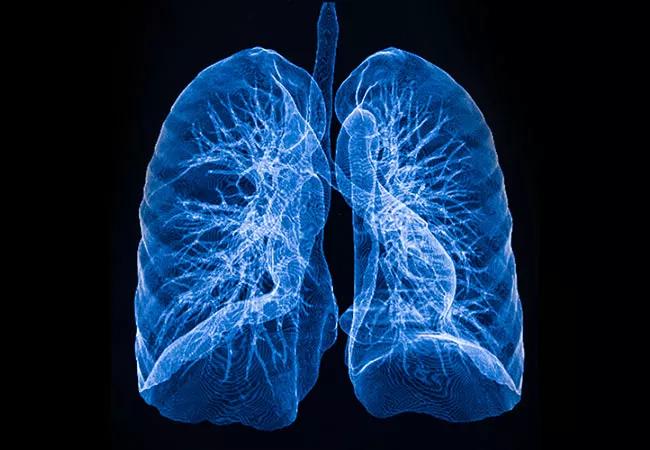
Decortication for lung transplant recipients with pleural space disease allows salvage of graft function with a reasonable morbidity profile. That’s the conclusion of a new retrospective analysis of more than 1,000 patients who underwent primary lung transplant at Cleveland Clinic.
Advertisement
Cleveland Clinic is a non-profit academic medical center. Advertising on our site helps support our mission. We do not endorse non-Cleveland Clinic products or services. Policy
“Decortication for pleural space management in appropriate patients, especially when performed early after transplantation, can help salvage pleural space and improve graft function,” says the study’s senior author, Usman Ahmad, MD, a thoracic surgeon at Cleveland Clinic. “The procedure was found to be acceptably safe, and it allowed these patients to achieve survival comparable to that among other primary lung transplant recipients.”
The study was published online ahead of print in the Journal of Heart and Lung Transplantation (2021 Mar 29;S1053-2498[21]02262-2).
Early and late pleural space complications commonly occur after pulmonary transplantation, sometimes involving mechanical restriction of the allograft. While thoracentesis helps achieve lung expansion, decortication may be necessary after recurrent pleural effusions, dense pleural adhesions or lung entrapment. However, reoperation on an allograft is associated with significant morbidity and mortality.
The indications, risk factors and outcomes of decortication following lung transplantation have not previously been well described.
The study analyzed 1,039 patients who underwent primary lung transplant at Cleveland Clinic between January 2006 and January 2017. Of these, 468 had pleural complications; 77 (16%) of these patients underwent 84 surgical decortication procedures for pleural space management. Most of these patients had already undergone a pleural intervention before decortication, most commonly thoracentesis and tube thoracostomy.
Advertisement
Indications for decortication included complex pleural effusions (56%), fibrothorax (20%), empyema (13%) and hemothorax (11%). Risk of decortication peaked at one month after transplant.
The following risk factors were identified for requiring decortication within the first month after transplant:
Patients with restrictive lung disease had a lower risk in this early phase. Factors increasing risk thereafter were diabetes and increased systolic pulmonary pressure.
Complications occurred after 30 procedures (36%), including prolonged (> 72 hours) intubation (10 patients), blood transfusion (9) and pulmonary complications (15). Postoperative respiratory failure occurred in 14 patients. Reintervention was necessary in 10 patients (2 thoracenteses, 1 tube thoracostomy, 7 reoperations).
After decortication, 88% of allografts had full expansion noted during surgery. Forced expiratory volume in one second (FEV1) increased 10 percentage points initially after decortication to a peak of 60%, followed by a slow decline to 55% by five years. Allograft function remained lower than that of patients without pleural complications, although the difference decreased over time.
No patient required repeat lung transplant after decortication, and survival was comparable to reported survival for all primary lung transplant recipients.
The study report also details management strategies informed by the Cleveland Clinic experience in these complex patients. The authors break down strategies into early- and late-phase pleural complications.
Advertisement
Early-phase loculated effusions are typically observed in the first few weeks after transplant, especially in patients who are fluid overloaded or have poor respiratory effort. For these patients, the following advice is offered:
Late-onset fibrothorax occurs in patients with declining lung function after the first month, with many having new-onset oxygen dependence. A dense peel, usually in the lower lobes, is typically seen on radiograph. Dr. Ahmad notes the following:
Advertisement
“We advise early intervention of loculated effusions to avoid late-onset fibrothorax, which is much harder to address,” concludes Dr. Ahmad. “Overall, despite its attendant morbidity, decortication allowed for management of pleural space pathology without further deterioration in allograft function, and it potentially prolongs time to graft failure.”
Advertisement
Advertisement

How Cleveland Clinic is using and testing TMVR systems and approaches

NIH-funded comparative trial will complete enrollment soon

How Cleveland Clinic is helping shape the evolution of M-TEER for secondary and primary MR

Optimal management requires an experienced center

Safety and efficacy are comparable to open repair across 2,600+ cases at Cleveland Clinic

Why and how Cleveland Clinic achieves repair in 99% of patients

Multimodal evaluations reveal more anatomic details to inform treatment

Insights on ex vivo lung perfusion, dual-organ transplant, cardiac comorbidities and more