Locations:

The rare and aggressive form of bone cancer requires specialized treatment, but new and emerging therapies are providing hope for patients
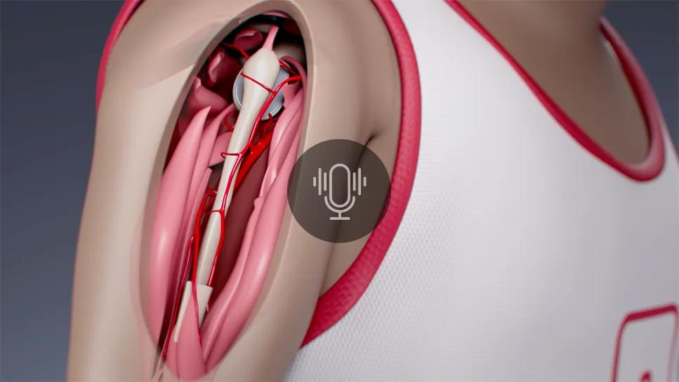
Image content: This image is available to view online.
View image online (https://assets.clevelandclinic.org/transform/8a46a619-1a2b-42d4-9333-537723574db8/cqd-osteosarcoma-cancer-advances-podcast2)
Illustration of osteosarcoma shoulder procedure
Sarcomas are among the rarest of cancer diagnoses, making up only about 1% of diagnoses in adults in the United States and ~15% of pediatric malignancies. Due to its rarity, there are not many medical centers that specialize in sarcoma treatment.
Advertisement
Cleveland Clinic is a non-profit academic medical center. Advertising on our site helps support our mission. We do not endorse non-Cleveland Clinic products or services. Policy
“A high-volume center may see 200-300 sarcomas in a year,” says Nathan Mesko, MD, Vice Chair of Global Clinical Operations for the Department of Orthopaedic Surgery and Center Director for Musculoskeletal Oncology. “Maintaining that expertise with a team that is really passionate about a rare cancer is an essential thing for the best possible outcomes, and it's one of the reasons why I love being here at Cleveland Clinic.”
Osteosarcomas — an aggressive bone cancer that generally affects teens and older adults — are often found around the knee, in the bottom part of the thighbone, the top part of the shinbone or in the shoulder.
“I think the ones that become even more problematic, just from a technical and complexity standpoint, are those that are in the spine or the pelvis,” says Dr. Mesko. “They become particularly challenging because of all the high-stakes anatomy in that area. But probably even more of a problem is that in the pelvis, a tumor can grow for a long time before it's noticed. These osteosarcomas will often be very large in size before they're identified. Sometimes patients don't develop symptoms until much later.”
In a recent episode of Cleveland Clinic’s Cancer Advances podcast, Dr. Mesko discusses some of the challenges associated with osteosarcomas and how Cleveland Clinic is reshaping outcomes through some innovative approaches to care.
Chemotherapy has long been the first line of treatment for osteosarcomas, and it remains in this position. Radiation, however, was not typically viewed as a treatment option. However, Dr. Mesko says this long-held belief is being challenged.
Advertisement
“We’re looking at ways that we can definitively treat metastatic sites and give people improved survival, better quality of life and function,” he explains. “We're thinking outside of the box with concepts and translating from other types of cancers, such as how do we boost the system with immunotherapy and make that synergistic with radiation? How do we extrapolate lessons learned from other cancers and translate them to osteosarcoma – an example being immunotherapy or radium, which was historically used with prostate cancer? We want to take these really difficult scenarios and give people hope.”
There have already been successes. Forty years ago, patients with osteosarcomas were almost automatically amputated. Now with chemotherapy, survival rates have increased, and amputation rates have dropped to ~5% of sarcoma cases.
Dr. Mesko also believes that chemotherapy helped surgical oncologists reduce the need for being so aggressive with their operations.
“The number one principle as a surgical oncologist is to get the cancer out,” he says. “With a bone sarcoma, you have to get clean margins. You have to remove normal, healthy tissue around the tumor to assure ourselves that the cancer is out. As we've advanced, we have a better understanding of the principles of chemotherapy and tumor necrosis and how that affects the risk of local recurrence. We can save blood vessels and nerves that we may not have otherwise saved and preserved function in that leg and balanced that oncologic and functional principle.”
Advertisement
Another result of this increased understanding is the ability to reconstruct. For example, if a patient had an osteosarcoma in the hip socket, they would previously have been left without a hip joint. A cadaveric bone could be used, but that increases the chance of a complication, and it could be very hard to construct and make sure the fit is correct. Now, technology has improved and evolved to the point that surgeons can be extremely precise in their geometric margins and create a “perfect match” implant that is created by a 3D printing technique – allowing us to tailor our approach individually to each patient.
“With 3D printers, we can build implants now that fit that exact congruency that we just created with those patient-specific cutting guides,” says Dr. Mesko. “We can get a perfect fit that reliably allows the bone to heal and gives us the ability to reconstruct anatomy where we can keep the limb lengths the same and provide patients with a high level of function. It's essentially like doing an ultra-fancy hip replacement, as an example, except in a way that nobody would have ever imagined 10 or 12 years ago.”
While 3D printing is something that can be employed in adults, it is not an option for pediatric cases since the patient is still growing.
“We had a pediatric patient a few years ago who was five years old,” says Dr. Mesko. “He was admitted to the hospital because his arm was hurting, and he had a big mass on his shoulder. It was an osteosarcoma. Routinely, in an adult, we can do a complex type of shoulder replacement and they’ll be functional. The trick with a five-year-old is that they’re still growing, and there isn’t anything I can easily pull out of the “usual bag of tricks” and say, ‘I’m going to rebuild your arm.’”
Advertisement
He continues, “You have a moment where you have to talk to mom and dad and say, ‘Well, the options are we can be really creative to a point where you might think I'm crazy. Or we can remove your five-year-old's arm. My number one priority is to get this cancer out of your son. If we don't, this is life-threatening. If we do, we give him a chance at a cure.’"
Dr. Mesko ended up using the patient’s fibula (small bone in the leg) to recreate the shoulder joint. Employing help from a plastic surgeon (Dr. Graham Schwarz), the multidisciplinary team removed the patient’s blood vessels from the top of the bone, and, under a microscope, then reattached them to the main blood supply of the arm.
“The beauty of something like that is twofold,” explains Dr. Mesko. “One, now I have a solution to actually give this kid an arm + hand + elbow that he can use. But two, if it works, the growth plate actually still continues to work and allows for growth. And that's one of the things we have to think about in kids is they're still growing. Adults are done growing, so I don't have to worry about how long this arm should be in five years or in 10 years. But in this patient, I had to. He had 10 years-plus of growth left and being able to borrow a bone that still gives me some of that growth was one of those ideas.”
When it comes to osteosarcoma care, Dr. Mesko says he is particularly excited about some of the emerging technologies on the horizon.
“A surgical oncology world with virtual reality and robotics has me salivating,” he says. “One of the hardest things is understanding where our margins are going to be. So, to be able to have 100% assurance that you're going to reproduce your plan each time, I think that has potential to drive margins to a point where a positive margin can be eradicated in some instances.”
Advertisement
Virtual reality represents another opportunity to push care forward. Although bones are fixed objects, muscles are not and can move during surgery. Dr. Mesko believes that virtual reality can be used to overlay anatomic structures to see exactly where a tumor or edema field is, which can improve their surgical planning.
Artificial intelligence also shows promise in this field. Studies have demonstrated that necrosis of the tumor is important in determining survival rates and whether the cancer comes back to the surgical field. Dr. Mesko explains that when there is more necrosis, he can cheat the margins and get closer to the tumor during surgery.
“The problem right now is that everything is after the fact,” he says. “I have to cut the tumor out, and then we look at the necrosis in everything. It doesn't help me plan or execute my surgery.”
But if a PET scan or an MRI could be used and analyzed by virtual augmentation or artificial intelligence, surgeons could drastically improve their decision-making.
Dr. Mesko explains, “What if we could actually say with a high degree of certainty, ‘Based on this MRI, you have “X” amount of necrosis.’ It really lets me go in with a much more confident understanding of the biology that I'm dealing with, going into the operating room. I think the sky is the limit when it comes to what technology can empower us to do in our field.”
Podcast content: This podcast is available to listen to online.
Listen to podcast online (https://www.buzzsprout.com/2241774/18742908)
Advertisement

Study also finds that 26% of children with cancer have mutations in DNA repair genes

Research highlights promising outcomes for treating recurrent and metastatic cases

Biologic approaches, growing implants and more

Innovative procedure offers less-invasive alternative to Latarjet procedure

Even patients with reported penicillin allergies can receive it without increased complications

Study challenges assumptions about risk evaluation in total hip revision

Protein expression in synovial fluid indicates patients’ immune factors may be involved

Cleveland Clinic’s Global Peak Performance Center and PGA TOUR partnership pair advanced assessment with longitudinal follow-up to enhance clinical decision-making