Anterior rectus sheath approach is key for keyhole technique
Cleveland Clinic is a non-profit academic medical center. Advertising on our site helps support our mission. We do not endorse non-Cleveland Clinic products or services. Policy
Minimally invasive open kidney transplants are safe, quick to perform and provide equivalent — if not better —outcomes compared with the conventional and essentially unchanged technique first described by French surgeon Rene Kuss, and refined and popularized by American surgeon Joseph Murray more than 60 years ago.
A 2-inch to 5-inch oblique skin incision about 2 inches above and parallel to the inguinal ligament is performed. This is carried down to the anterior rectus sheath, which is divided longitudinally. The rectus muscle is then retracted medially, in the process exposing the inferior epigastric vessels and the spermatic cord/round ligament.
Figure 1. Skin and anterior rectus sheath incisions.
Figure 2. Rectus sheath incision with medial retraction of rectus muscle and exposure of the cord structures, inferior epigastric vessels, transversalis fascia and peritoneum.
Figure 3. Ligation and division of inferior epigastric vessels, opening of the transversalis fascia, medial retraction of the peritoneum, exposure of the external iliac vessels and development of the iliac fossa pocket for the kidney.
Figure 4. Implanted kidney in final position.
Access into the retroperitoneum is achieved by dissecting extraperitoneally between the transversalis fascia and the peritoneum, inferior to the arcuate line (linea semicircularis of Douglas). A pocket is then created for the allograft.
After adequate exposure of the external iliac vessels, the allograft is implanted. Vascular anastomosis may be performed extracorporeally or in situ (with the kidney in its final position) or may be done in combination, mostly depending on the recipient’s anatomy. Lich ureteroneocystostomy is then accomplished as usual. The anterior rectus is closed using absorbable monofilament, the subcutaneous tissue is reapproximated to eliminate dead space and the skin is closed subcuticularly. No drains are used.
This approach ensures a faster, better and stronger musculofascial layer closure than the conventional approach, which is done through the external oblique, internal oblique and transversus abdominis muscles/aponeuroses in one, two or sometimes three layers. Due to the fact that muscles are used to hold sutures in the standard technique, these may rip and result in wound dehiscence or hernia.
Additionally, the peritoneum is at risk of being caught with the suture during closing in the standard procedure. This is avoided in the anterior rectus sheath approach because the rectus muscle protects the peritoneum during suturing.
In the keyhole technique, minimal dissection is carried out — only what is necessary to fit an allograft and expose the external iliac vessels and bladder. Wound complications are practically eliminated and the total operative time is shortened significantly (an average of two hours versus three to four hours for traditional surgery) as opening, exposure and closing times are minimized.
Compared with recently reported laparoscopic and robotic transplants, this open procedure is less costly and significantly faster to perform (laparoscopic procedures may take an experienced surgeon four to six hours to perform; robotic procedures take four to eight hours). More important, ischemia time is shorter, thus not compromising immediate allograft function.
The length of the incision is about the same as in laparoscopic and robotic procedures, determined only by the size of the allograft. The allograft remains extraperitoneal, avoiding complications peculiar to intraperitoneal procedures. Finally, experienced transplant surgeons can easily adopt this technique without the costly and risky learning curves associated with high-tech procedures.
Minimally invasive surgeries cause minimal skin, muscle and tissue damage, which means less pain, less scarring, quicker recovery, fewer wound complications and shorter hospital stays. In kidney transplant surgery, these benefits are magnified.
Interestingly, minimal open techniques have recently been described but have not been widely adopted. A simple, small-incision technique — without the help of laparoscopic or robotic instruments — using the anterior rectus sheath approach described above results in excellent renal transplant operative outcomes.
Dr. Africa is a staff physician in Cleveland Clinic’s Glickman Urological & Kidney Institute, practicing in the Charleston, W. Va., Urology Office.
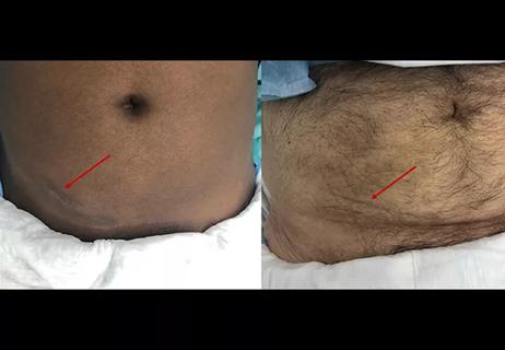
Smaller incision may lead to reduced postoperative pain for some patients
Improving access to lifesaving kidney transplant
The process could improve access and equity for patients with end-stage kidney disease
Video offers glimpse into technically challenging portion of the procedure
Infectious disease and kidney transplant specialists share key insights
Demographic and social factors found to influence preemptive listing
New interventions and policies haven’t increased wait-listing
Cleveland Clinic surgeons use single-incision robotic surgery to perform kidney transplant