Locations:

Innovative care sustains baby with complex heart disease
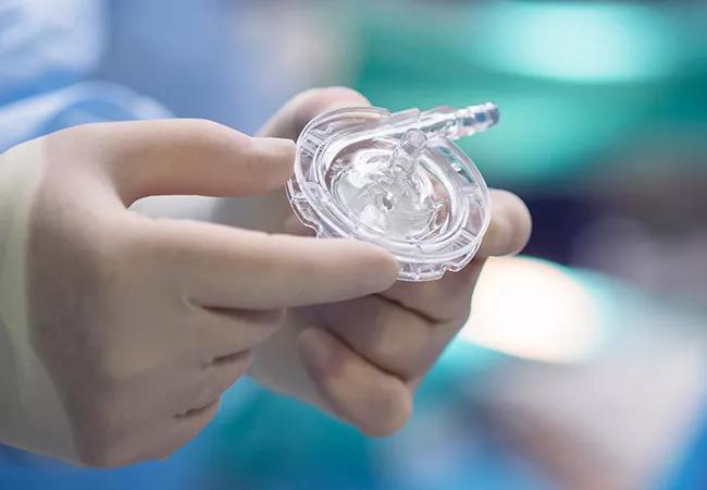
Image content: This image is available to view online.
View image online (https://assets.clevelandclinic.org/transform/2630791c-59a4-427a-b8d4-6da5efc1eb38/650x450-PediMag-ventricular-assist-device_jpg)
650×450-PediMag-ventricular-assist-device
By Gerard Boyle, MD, and Hani Najm, MD
Advertisement
Cleveland Clinic is a non-profit academic medical center. Advertising on our site helps support our mission. We do not endorse non-Cleveland Clinic products or services. Policy
A bridge to transplantation can require as much or more clinical innovation than the transplantation itself.
Such was the case with one newborn at Cleveland Clinic who had a complex form of congenital heart disease, making conventional repair impossible and survival until transplantation unlikely. In this case study, we discuss the patient’s months-long wait for a heart transplant and the innovative care that kept her alive.
The patient was born with pulmonary atresia with an intact septum (hypoplastic right ventricle). She had patent ductus arteriosus (PDA), a duct between her aorta and pulmonary artery that supplied her lungs.
Prostaglandin initially helped keep the ductus open while the patient awaited a permanent repair. However, a BT shunt became necessary to maintain the flow of blood to her lungs longer term.
In addition to these heart defects, the patient also had right ventricle-dependent coronary circulation. Her heart was supplied not by normal coronary arteries but by connections inside her (abnormally small) right ventricle.
Following the insertion of the BT shunt, and because of her dependency on blood from the right ventricle, she was not able to sustain good ventricular function. She was placed on ECMO and listed for heart transplant.
We feared that ECMO would not keep her alive long enough to wait for a transplant — or that she would not be weanable from it due to her right ventricle dependency. ECMO would suffice for only one week to 10 days, certainly no more than two weeks.
Advertisement
Knowing that the patient’s lungs were normal, we needed to replace only her ventricular function. We put her on a long-term ventricular assist device (VAD), an extracorporeal centrifugal pump without an oxygenator.
Because she had duct-dependent circulation, we attached the VAD by injecting into the descending aorta, beyond the ductus arteriosus. This prevented less-oxygenated blood from the VAD being pumped into the aorta. By bypassing the ductus, her heart continued to pump higher-oxygenated blood to itself and her brain, while the VAD pumped less-oxygenated blood to the rest of her body.
This was acceptable because a newborn’s lower body can live with less oxygen. Our solution allowed us to maintain an oxygen saturation of 90 percent to the patient’s upper body and a saturation of 50 to 60 percent to the lower body.
Significant anticoagulation is required for patients on this type of VAD. Heparin is standard, yet it is not without concern when used in infants.
Frequent partial thromboplastin time (PTT) tests are required — several per day. Each test requires a large volume of blood, and recurring tests often necessitate blood transfusion. Blood transfusion can create complications for patients preparing for transplant as exposure to transfusions can alert the infants immune system (sensitize it) to possible donor organs and thereby increase the risk of rejection.
In addition, for unknown reasons, the PTT test is often unreliable in infants. It is not uncommon to see blood clots in the VAD of patients whom we thought were sufficiently anticoagulated. However, the PTT test is somehow more reliable for patients taking bivalirudin. More reliability means fewer tests are needed, which means less blood loss and fewer transfusions.
Advertisement
For these reasons, the patient received bivalirudin, instead of heparin, which kept her sufficiently anticoagulated with no harmful effects. This approach is relatively new and is just beginning to be acknowledged at other medical centers.
At three months of age, the patient had a heart transplant.
Now at 13 months, she has recovered all organ function, although she has a tracheostomy and remains on a ventilator. She has begun breathing on her own for several hours per day and soon will have the tracheostomy removed. No transplant rejection has been detected. She has started to stand and appears to be developing normally. She is quite social, interactive and playful.
This is a remarkable outcome given her degree of illness. Survival to transplant had been unlikely, yet several novel treatments helped us overcome the complexity of her condition and sustain her long enough to locate an organ match.
With modified techniques, it is feasible to employ VAD long-term in an infant with right ventricle-dependent coronary circulation and duct dependency.
Dr. Boyle is Medical Director of Pediatric Heart Failure and Transplant Services at Cleveland Clinic. Dr. Najm is Chair of Pediatric and Congenital Heart Surgery at Cleveland Clinic.
Advertisement
Advertisement

A snapshot of key stats from one of the world's busiest centers

‘Sac flow’ is more precise and will ease unfounded patient concerns, experts argue

Join us in New York Dec. 4-5 for evidence-based instruction with real-world examples

First-ever transcarotid artery revascularization trial with no strokes or device-related deaths

Consensus statement outlines the team, infrastructure and experience needed to deliver TTVI safely and effectively

Innovative approach to living-tissue AVR achieves low reintervention rates, excellent long-term survival

Diagnosis and treatment of malnutrition and cachexia are key to improving cardiac outcomes

Symptom burden at presentation is a potent predictor of long-term survival, large analysis shows