Locations:

Complex chondrosarcoma cases pose challenges, utilize partnership
As an orthopaedic oncologist, Nathan Mesko, MD, partners with specialists across the enterprise, “crossing the usual boundaries of extremity surgery,” he says.
Advertisement
Cleveland Clinic is a non-profit academic medical center. Advertising on our site helps support our mission. We do not endorse non-Cleveland Clinic products or services. Policy
This was true recently when Dr. Mesko partnered with thoracic surgeon Daniel Raymond, MD, of Cleveland Clinic’s Department of Thoracic and Cardiovascular Surgery, on two separate chondrosarcoma cases. Chondrosarcoma, a rare and malignant primary bone sarcoma that develops in cartilage cells, can be difficult to diagnose and treat. Tumor resection is the preferred management of the disease.
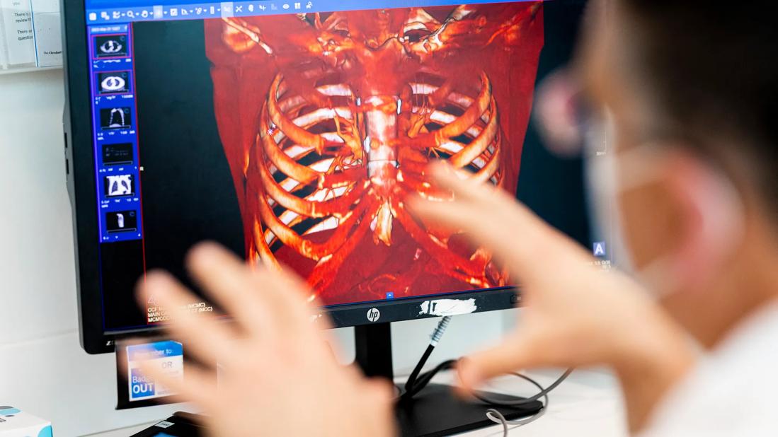
The first case involved a man in his mid-20s, who presented with chondrosarcoma of the manubrium, the top part of the breastbone. It was a low-grade tumor, Dr. Mesko explains. The goal was to remove it cleanly, sparing as much anatomy as possible.
By working with industry partners, the team was able to create a custom cutting jig design, allowing them to gain accurate margins while sparing as much bone as possible for reconstruction purposes. Next, they reconstructed the defect, utilizing orthopedic techniques, involving screws and bone cement, to create a rebar and wet concrete-type reconstruction of the manubrium. This approach enabled the patient’s full function immediately.
The second case involved a man in his 70s, who had undergone multiple resections of sternal chondrosarcoma. Dr. Mesko and his team were tasked with removing about 70% of his sternum bone and a portion of the medial clavicle while navigating precarious anatomic architecture behind the sternum.
“With such a large defect, leaving the defect open would predispose the patient to direct trauma to his heart and progressive spinal deformity, both of which, ultimately, would be life-threatening,” he says.
Advertisement
The team created a 3D model to prepare for the resection by working with the local bone bank and industry partners. They used measurements from the anticipated defect in the chest and identified a matching femoral donor bone allograft, utilizing a cadaveric femur to reconstruct this defect.
Because of the limited bone fixation, the team utilized multiple orthopaedic principles to help gain fixation, including plating, long cannulated screws and suspension with strong suture material. The graft was also supplemented with bone cement to increase its strength and all of the junctions were bone grafted.
Video content: This video is available to watch online.
View video online (https://www.youtube.com/embed/3oQNX3AcsbE?feature=oembed)
Teamwork: Chondrosarcoma of the Manubrium and a Sternal Chondrasarcoma (Graphic)
Dr. Mesko reinforces the importance of multidisciplinary environments that facilitate collaboration between seemingly disparate specialties. “There is no such thing as lone cowboys in the world of complex cancer surgery,” he remarks.
“My partners are my lifeline and my fuel, and being able to tell a patient that we got your cancer out and the surgery went off without a hitch can only occur when you have partners that are united and invested in the common goal.”
Advertisement
Advertisement

How it’s similar but different from the direct anterior approach

Collaboration must cross borders and disciplines

Systematic review of MOON cohorts demonstrates a need for sex-specific rehab protocols

Should surgeons forgo posterior and lateral approaches?

How chiropractors can reduce unnecessary imaging, lower costs and ease the burden on primary care clinicians

Why shifting away from delayed repairs in high-risk athletes could prevent long-term instability and improve outcomes

Multidisciplinary care can make arthroplasty a safe option even for patients with low ejection fraction

Percutaneous stabilization can increase mobility without disrupting cancer treatment