Locations:





AI tool matches manual grading and may improve consistency in retinal research
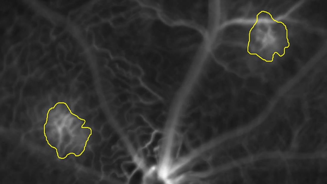
Image content: This image is available to view online.
View image online (https://assets.clevelandclinic.org/transform/f7bdf86b-2db0-458d-84df-f7af97b3ec09/choroidal-neovascularization-fluorescein-angiography-feature)
Yellow outlines on two areas of white lines on a black background
For ophthalmic researchers developing therapies for choroidal neovascularization (CNV), a critical step can require hours of manually tracing blurry, indistinct boundaries of CNV lesions on fluorescein angiography (FA) images. The process is tedious, subjective and can introduce grader-to-grader variability, potentially complicating preclinical drug evaluation and slowing drug development.
Advertisement
Cleveland Clinic is a non-profit academic medical center. Advertising on our site helps support our mission. We do not endorse non-Cleveland Clinic products or services. Policy
Now, Cleveland Clinic Cole Eye Institute is developing a deep learning-based system that automates the process. The technology could help researchers evaluate new therapies more quickly and consistently, bringing greater efficiency and reproducibility to preclinical research.
“Quantification of CNV lesions is essential for evaluating the efficacy of experimental therapies for neovascular age-related macular degeneration and other diseases,” says Rupesh Singh, PhD, Director of the In Vivo Imaging Core Laboratory at the Cole Eye Institute. “An automated approach can achieve strong agreement with expert manual annotations while being faster and more consistent across studies.”
At the 2026 ARVO Annual Meeting, Dr. Singh presented how he and his team created an artificial intelligence (AI)-based system trained on FA images with CNV lesions outlined by manual graders. The tool, DeepLabV3+, learned to classify each pixel as either lesion or background, enabling precise border identification and automated calculation of lesion area.
Next, the team tested the system in two preclinical models, one with laser-induced CNV and another with spontaneous multifocal CNV.
“We deliberately tested the algorithm in two distinct models to determine whether it could handle different lesion phenotypes and morphologies,” Dr. Singh says. “One model produced laser-induced lesions, which were more localized and focal, while the other model generated spontaneous lesions of varying size and morphology. A useful AI tool must be able to quantify a broad range of lesion patterns, not just those it encounters most frequently during training.”
Advertisement
Compared with manually annotated reference masks, the AI tool demonstrated strong segmentation performance in both preclinical models. Among other metrics, the team calculated:
“These scores show the algorithm does more than simply flag lesions,” Dr. Singh says. “It precisely measures lesion boundaries and reliably distinguishes disease from normal tissue. Perhaps most importantly, it maintains this accuracy in the more complex, multifocal genetic model, suggesting robustness across two distinct preclinical CNV phenotypes.”
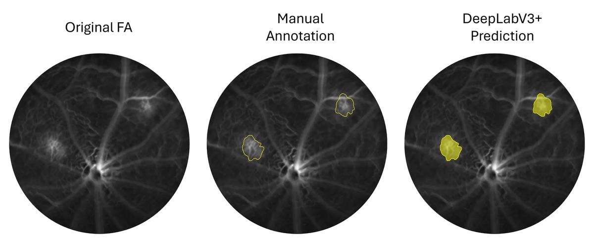
Image content: This image is available to view online.
View image online (https://assets.clevelandclinic.org/transform/d30730e7-d400-4da2-91b7-92733c3bf5c6/choroidal-neovascularization-fluorescein-angiography-inset)
Fluorescein angiography (FA) image demonstrating choroidal neovascularization (CNV) segmentation. Left: original FA image. Middle: manually delineated lesion boundaries. Right: lesion segmentation predicted by DeepLabV3+.
Now validated in two preclinical CNV models, this AI tool has the potential to standardize CNV quantification in experimental drug testing, streamline image analysis and reduce intergrader variability.
For longitudinal studies — where consistency across time points and graders is critical — automated quantification could significantly boost the reliability and speed of data reporting.
Advertisement
Although adaptation for human retinal imaging would require retraining and validation on larger, more heterogeneous human datasets, the technology itself is transferable, Dr. Singh says.
“Human CNV lesions are far more variable in size, severity and treatment status than those in preclinical models,” he says. “Retraining the algorithm will demand multicenter data and rigorous standardization, but the prospects are promising.”
Beyond FA, the system could be extended to other imaging modalities, including OCT, OCT angiography and fundus autofluorescence, enhancing disease progression assessment.
“We have already used deep learning with OCT imaging to identify disorganization of retinal inner layers, a biomarker in diabetic retinopathy,” Dr. Singh says. “That experience shows how similar AI approaches can be adapted to different retinal imaging modalities and disease biomarkers. These methods can ultimately provide a more comprehensive and objective analysis of retinal disease.”
Advertisement
Advertisement

New studies clarify how sensory loss and nystagmus shape real-world visual function
Quantitative B-scan findings may offer a noninvasive way to assess vascular inflammation
Learn the latest recommendations in diagnosis, treatment and surveillance
The drug does not provide lasting disease control for most patients
A review of pathophysiology, risk factors, clinical presentation and treatment options

Registry data highlight visual gains in patients with legal blindness
Study is first to show reduction in autoimmune disease with the common diabetes and obesity drugs
The advanced stage of diabetic retinopathy is among the most challenging for retinal surgeons
 Search