Locations:

Optimally timed valve replacement depends on an expert approach to nuanced presentations
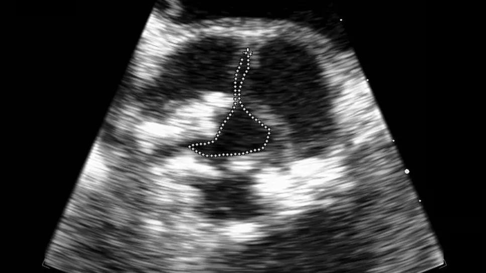
Image content: This image is available to view online.
View image online (https://assets.clevelandclinic.org/transform/8b023293-7141-4b76-b47d-9218ac56fdce/aortic-stenosis-echocardiogram-feature)
echocardiogram showing severe aortic stenosis
When the American College of Cardiology (ACC) and the American Heart Association (AHA) issued their latest guideline on the management of valvular heart disease four years ago, it reflected what was then relatively recent consensus on when and in whom aortic valve replacement (AVR) should be considered for treatment of valvular aortic stenosis (AS). That consensus has only solidified in the subsequent years, but there remain important nuances in the evaluation of patients with AS that can be difficult for guidelines to elucidate.
Advertisement
Cleveland Clinic is a non-profit academic medical center. Advertising on our site helps support our mission. We do not endorse non-Cleveland Clinic products or services. Policy
“The evaluation and diagnosis of AS may be perceived as fairly straightforward, but it often tends to get complicated, especially when one factors in the question of exactly when to intervene,” says Milind Desai, MD, MBA, Medical Director of Cleveland Clinic’s Aorta Center and a cardiologist in its Valve Center.
The ACC/AHA guideline (Circulation. 2021;143[5]:e35-e71) identifies four broad categories of indications for intervention for AS with AVR, whether surgical or transcatheter:
In real-world practice, however, these broad indications can quickly get clouded by discrepancies between AS severity and the patient’s reported symptoms, between aortic valve area and mean pressure gradient, and between findings from different imaging modalities (Figure 1). “Patients often meet some criteria for severe AS but not all the criteria,” notes Brian Griffin, MD, Head of Cleveland Clinic’s Section of Cardiovascular Imaging and Medical Director of its Valve Center.

Image content: This image is available to view online.
View image online (https://assets.clevelandclinic.org/transform/81a60058-facd-4eba-b382-ca0b1c5b0a8c/aortic-stenosis-echocardiogram-inset1)
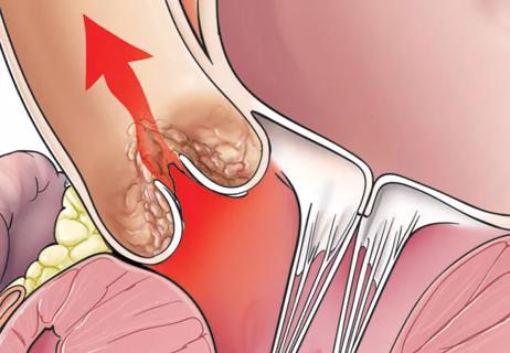
Figure 1. Schematic illustrations contrasting a normal, stenosis-free aortic valve with low-flow, low-gradient aortic stenosis (AS). These scenarios show why normal left ventricular ejection fraction (LVEF) does not always generate a high gradient in AS. LVH = left ventricular hypertrophy, with “adequate LVH” signifying an adequate degree of hypertrophy to maintain normal wall stress and “inadequate LVH” signifying hypertrophy insufficient to normalize wall stress, leading to afterload mismatch and progressive dilatation of the left ventricle with reduced function.
“About a quarter of patients with AS seen at Cleveland Clinic have normal LV function with a valve area and pressure gradients that are not fully in sync in terms of stenosis severity,” Dr. Griffin continues. “In those cases we have to make a judgment whether or not it’s true stenosis.” He adds that another 5% of patients have low gradients, a severely reduced valve area and severely reduced LV function. “That amounts to about 30% of patients in whom disease classification and optimal management are difficult to figure out.”
Advertisement
Dr. Desai says some of the most common nonstandard presentations include the following:
“Separating these presentations from standard severe AS can be daunting, and it has implications for appropriate timing of intervention,” Dr. Desai observes.
The challenge is overcome with various forms of additional testing beyond initial echocardiography. Often that means multimodality imaging, most often with the addition of CT. “Most of our patients nowadays undergo CT along with echo,” Dr. Desai says. “If CT cannot be performed, MRI, transesophageal echo [Figure 2] or 3D echo can help us get a good handle on the valve area or identify something we might be missing on standard echo.”

Image content: This image is available to view online.
View image online (https://assets.clevelandclinic.org/transform/f262bf93-cb91-4876-977d-86272619719d/aortic-stenosis-echocardiogram-inset2)
Figure 2. Severe aortic stenosis planimetry on transesophageal echocardiography.
CT also is integral to another type of additional testing that can help define AS severity: calcium scoring of the aortic valve. “When necessary, we use CT scanning to assess the amount of calcium on the valve,” Dr. Griffin explains. “Scoring is a bit different for women versus men, but in either sex the presence of a high calcium burden on the valve makes severe AS highly likely, even if the patient doesn’t meet all the echo criteria. We do a lot of calcium scoring to make sure referrals for early intervention are appropriate.”
Advertisement
Exercise stress echocardiography can be another helpful indicator of increased risk from diminished functional capacity due to asymptomatic AS. Recognition of its role in this setting stems in part from a large Cleveland Clinic analysis showing improved survival following AVR in asymptomatic patients with AS undergoing stress echo (Circ Cardiovasc Imaging. 2016;9[7]:e004689)[GC1] . “We now use stress echo quite a bit when trying to decide the timing of intervention, especially when patients report no symptoms but we suspect their AS is getting worse based on other findings,” Dr. Griffin says.
Biomarkers such as B-type natriuretic peptide (BNP) can also be useful in that context. “When a patient has severe AS but is asymptomatic, if their BNP level is two or three times normal, then valve replacement can and should be considered,” Dr. Griffin notes.
Indeed, Drs. Desai and Griffin say a resourceful, refined testing and evaluation strategy can make the biggest difference for patients who perceive themselves as asymptomatic. “We are not interested in sending patients for unnecessary procedures,” Dr. Desai explains, “but when we have findings that strongly suggest severe AS despite the patient reporting no symptoms, our approach is to thoroughly assess whether the patient may in fact be able to benefit from intervention. That can be done with a stress test or strain imaging to confirm whether they are indeed asymptomatic or by calcium scoring, blood testing or additional imaging to make sure nothing is being missed.”
Advertisement
The impetus for such thoroughness is the benefit that’s been demonstrated from timely AVR. “Patients develop structural changes in their heart related to AS,” Dr. Griffin says. “Resulting thickened muscle may regress after the valve is replaced, but it doesn’t always regress completely to normal. That leaves patients at risk for a stiffer heart that’s more likely to develop heart failure and other complications. Data show that earlier intervention in severe AS extends patients’ survival and quality of life.”
The benefits of early intervention are likely to be greatest at high-volume centers with very low rates of mortality and other complications, an observation made in the latest ACC/AHA guideline. “At a place like Cleveland Clinic, where procedural mortality rates for AVR are consistently 0.5% or less, the case for early intervention for AS is particularly compelling,” Dr. Desai concludes.
Advertisement
Advertisement

JACC State-of-the-Art Review details current knowledge and new developments

Age and other factors figure into the choice among SAVR, TAVR, Ross, Ozaki and more
Large longitudinal study supports earlier intervention over clinical surveillance

‘Sac flow’ is more precise and will ease unfounded patient concerns, experts argue

Innovative approach to living-tissue AVR achieves low reintervention rates, excellent long-term survival

Robust signal from observational study raises prospect of a long-sought medical therapy

Expert advice on repair vs. replacement, timing of surgery in asymptomatic cases and much more

Modified-Bentall single-patch Konno enlargement (BeSPoKE) optimizes hemodynamics, facilitates future TAVR