Locations:


New review proposes a multimodality framework for matching patients to procedures
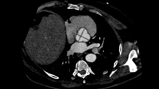
Image content: This image is available to view online.
View image online (https://assets.clevelandclinic.org/transform/a83ae412-4dfe-46fd-bcc0-90da9c17127b/Aortic-Ratio-CQD-hero)
axial image of the human chest showing the heart and other organs
With the plethora of surgical and transcatheter options now used to treat aortic valve (AV) and aortic root disease, carefully tailoring interventions to each patient’s anatomic and clinical features and long-term needs is essential. Fundamental to decision-making is thorough assessment with multimodality imaging, including echocardiography, computed tomography (CT), cardiac magnetic resonance (CMR) and, in some cases, nuclear imaging.
Advertisement
Cleveland Clinic is a non-profit academic medical center. Advertising on our site helps support our mission. We do not endorse non-Cleveland Clinic products or services. Policy
A recently published review article in Circulation: Cardiovascular Imaging proposes a multimodality imaging framework for optimizing AV and root procedures. Authored by Cleveland Clinic cardiovascular imaging specialists and cardiac surgeons, the review features medical illustrations, succinct tables and a case study along with detailed text to provide an overview of the distinct roles of various imaging methods for AV and aortic root procedures.
“We have more choices than ever before to address a patient’s individual needs, but we need to carefully consider the trade-offs between immediate risk, durability and options for future reintervention,” says senior and corresponding author Milind Desai, MD, MBA, Co-Director of Cleveland Clinic’s Cardiovascular Imaging Laboratories. “This review provides wide and deep guidance on matching patients with procedures across the spectrum of AV and aortic root disease.”
For AV disease, echocardiography reveals lesion severity, ventricular response and associated aortopathy. An essential study for aortic regurgitation, aortic stenosis, mixed aortic disease and valve disease with aortopathy, echo enhances hemodynamic phenotyping, mechanism definition and determining procedural relevance.
Three-dimensional transesophageal echocardiography (TEE) adds an important tool for assessing valve leaflet features and evaluating left ventricular (LV) global longitudinal strain.
“Echocardiography remains the basis for evaluating AV disease,” Dr. Desai says. “CT and CMR are key adjuncts when more information is needed on anatomic and volumetric features.”
Advertisement
CT provides detailed anatomic evaluation of the AV-aortic root complex and thoracic aorta, often improving on information provided by echocardiography. CT is the better tool for obtaining annular measurements for prosthesis sizing.
CT also best provides detailed calcification assessment, especially important in aortic stenosis and aortic regurgitation and for deciding between valve replacement and repair.
For chronic aortic regurgitation, CMR is an excellent tool to determine regurgitant fraction and LV volumes. Phase-contrast imaging is especially useful in patients with eccentric or multiple jets. Late gadolinium enhancement and parametric mapping can detect myocardial fibrosis. CMR also reveals details of aortic wall changes seen in inflammatory states.
Especially for younger patients, CMR can play an important role in aortic surveillance, as it reduces ionizing radiation exposure compared with serial CT.
Nuclear imaging, primarily positron emission tomography, is mainly useful for suspected inflammation or infection. It can help diagnose and monitor aortitis and improve detection of active infection of a prosthetic valve or graft.
“For different aortic valve and aortic root disease manifestations, integrating information from multiple imaging techniques helps determine smarter strategies based on knowledge beyond a patient’s diagnosis,” says Susan Keen, MD, first author of the review article and a cardiologist in Cleveland Clinic’s Section of Cardiovascular Imaging. “We are now well positioned to more thoroughly understand individual anatomic and physiological features with the goal of optimizing lifelong management.”
Advertisement
Multimodality imaging assists in deciding between use of a mechanical or a biological valve — as well as in avoiding prosthesis-patient mismatch — beyond the conventional factors of age, sex, body surface area, anticoagulation tolerance and comorbidities. For a small orifice, a mechanical valve may be indicated to optimize flow and long-term gradients. Preoperative CT or 3D TEE provides annular and sinus estimates that help predict the need for annulus enlargement. Indicators of increased risk of patient-prosthesis mismatch include smaller aortic dimensions on echo and a smaller sinutubular junction on CT.
Imaging can help predict durable AV repair by assessing leaflet tissue quality, lesion mechanism and annular geometry. Intrinsic leaflet disease or multifactorial lesion mechanisms predict poorer outcomes. If the underlying annular-root complex is abnormal, durable repair is rarely achieved from leaflet correction alone.
Multimodality imaging is important for helping determine whether a transcatheter approach is reasonable by defining annular-root anatomy, calcification extent, coronary access and existing aortopathy. Especially for young patients, multimodality imaging is invaluable for informing potential future interventions, such as valve-in-valve feasibility and surgical or transcatheter procedures.
TEE is an excellent tool for assessing key elements predicting success, such as cusp mobility, tissue quality and mechanism of regurgitation (geometric vs. degenerative). CT is important for defining root morphology and the extent of aortopathy.
Advertisement
A successful Ross procedure requires careful preoperative imaging assessment. Initial echocardiographic morphologic assessment of the aortic and pulmonary valves is critical. CT adds to the assessment of the annulus-root-ascending aorta complex.
“With more patients enjoying long-term survival following aortic valve and root surgeries, we are increasingly seeing the need for reinterventions,” says cardiothoracic surgeon Lars Svensson, MD, PhD, co-author of the paper and Chief of Cleveland Clinic’s Heart, Vascular & Thoracic Institute. “Multimodality imaging plays an important role in guiding strategies by helping determine whether reintervention is feasible and supports postoperative clinical benefit.”
He adds that determining the mechanism of failure is critical, as success is more likely if imaging shows a discrete and correctable problem. TEE is usually the first step, often followed by CT to clarify the anatomy and spatial relationships to help determine reentry risk and technical feasibility. CMR can provide additional information by quantifying regurgitation and assessing ventricular remodeling.
Advertisement
Advertisement
Earlier detection is reshaping timing and safety of these increasingly common surgeries

Elevated levels of the gut microbial metabolite predict prevalence and adverse outcomes in community-based study
Large analysis adds to evidence that absolute diameter alone may miss vulnerable patients
Height-indexed aortic measurements may better identify risk than diameter alone
Robust signal from observational study raises prospect of a long-sought medical therapy
New Cleveland Clinic data challenge traditional size thresholds for surgical intervention
Study finds comparable midterm safety outcomes, suggesting anatomy and surgeon preference should drive choice
New review distills insights and best practices from a high-volume center
 Search