Locations:

Technique features heavyweight guidewire and electrocautery to access the aortic sac
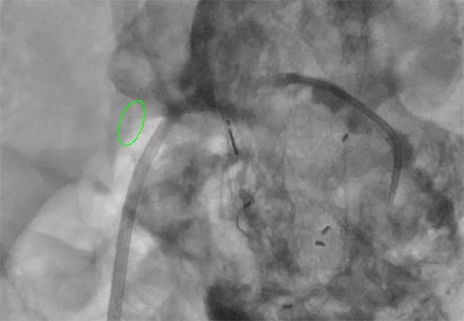
Image content: This image is available to view online.
View image online (https://assets.clevelandclinic.org/transform/a25c685d-5205-436d-be0f-c080636d97b8/23-HVI-4048160_type-II-endoleak_650x450_jpg)
imaging study of type II endoleak
For embolization of type II endoleak after endovascular aneurysm repair, use of a 0.014” guidewire and continuous current electrocautery allows crossing of the inferior vena cava/aortic sac junction without puncturing or damaging the endograft or adjacent structures. Cleveland Clinic vascular surgeons reported their experience with this novel technique in 12 patients in a retrospective cohort study published in Annals of Vascular Surgery (2023;93:300-307).
Advertisement
Cleveland Clinic is a non-profit academic medical center. Advertising on our site helps support our mission. We do not endorse non-Cleveland Clinic products or services. Policy
“The big advantage of this method is the sole use of a 0.014” guidewire to gain access to the aortic sac, making risk of inadvertent perforation of the endograft very unlikely,” says senior author Sean Lyden, MD, Chair of Vascular Surgery at Cleveland Clinic. “In all cases, access was successful without significant acute morbidity or mortality.”
Type II endoleaks ― i.e., flow into and out of the aneurysm sac from a patent lumbar and/or inferior mesenteric artery ― sometimes develop after endovascular aortic aneurysm repair (EVAR). Because they are common and rarely rupture, intervention is recommended only if the aortic sac expands by 5 mm or more. With the goal of saving the endograft without needing to explant the original EVAR, embolization or ligation of the feeding vessels should be undertaken.
Transarterial, translumbar and transgraft approaches have been developed to access the aortic sac for embolization. A transcaval approach is increasingly used when translumbar or transarterial access has failed or is not feasible. But inconsistent access and needle injury to the intact endograft have been reported. Most published methods describe using a 14- or 16-gauge transhepatic jugular needle to create the aortocaval crossing.
To reduce the access challenges and risk of needle injury, Cleveland Clinic vascular surgeons developed their transcaval technique using an electrified 0.014” chronic total occlusion wire to enter the aortic sac. Their Annals of Vascular Surgery publication details the technique in addition to reporting outcomes in the initial patients undergoing the procedure.
Advertisement
Transcaval embolization is performed in a hybrid operating room. A DynaCT scan is used to fuse the preoperative CT imaging to the patient’s anatomy to mark the location of intended crossing from the inferior vena cava into the aortic sac.
The procedure is done via percutaneous femoral venous access, allowing the patient to remain supine using local anesthetic. A steerable sheath and catheter are used to direct a 0.014” heavyweight tip wire guide to the area of crossing, and continuous current electrocautery is used to create the connection between the inferior vena cava and the aortic aneurysm sac. Selective embolization of the endoleak outflow vessel is undertaken using coils or liquid embolic agents. For cases in which selective embolization is not possible, the aneurysm sac is filled with hemostatic agents to induce occlusion. Most patients are treated on an outpatient basis with same-day discharge. Key stages in the procedure are depicted in the Figure.
Dr. Lyden highlights a couple of technical tips:
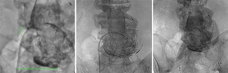
Image content: This image is available to view online.
View image online (https://assets.clevelandclinic.org/transform/23e30de5-cf24-4b15-93ce-e7a2274104f7/23-HVI-4048160_transcaval-endoleak-embolization_jpg)
Figure. Left: DynaCT scan with circle identifying the location for crossing into the aortic sac. Contrast is injected by catheter to demonstrate successful crossing. Middle: Image showing transcaval access with a 6.5F steerable sheath advanced into the aneurysm sac. Right: The sac filled with liquid embolic after treatment.
Twelve patients (83% men, average age [± SD] 79 ± 6 years) were treated with the novel transcaval access approach between March 2019 and June 2021. Overall, they had a high number of comorbidities, including essential hypertension, hyperlipidemia, coronary artery disease and chronic obstructive pulmonary disease.
Advertisement
All patients had type II endoleak with a documented aneurysmal sac diameter increase of at least 5 mm and an average aneurysm diameter of 7.8 ± 2.2 cm. Five patients had previous transarterial or translumbar embolization. The median time between initial EVAR implantation and transcaval embolization was 51.4 months (range, 1.5-113.1).
Median follow-up after transcaval embolization in the 12 patients was 6.5 months (range, 0-14.8). Procedural and follow-up outcomes included the following:
Advertisement
Drs. Lyden and Caputo say that they choose the approach to address type II endoleak by considering the source of the endoleak and the expected difficulty in directly accessing the outflow source. “For inferior mesenteric artery endoleaks, we prefer the transarterial approach,” Dr. Caputo says. “Translumbar access is preferred when there is a large lumbar artery not adjacent to the endograft and a clear access window exists.”
The surgeons note that the transcaval approach is limited by the difficulty of steering catheters and microcatheters into the outflow vessels, resulting in persistent endoleak in some cases.
“However, we are increasingly using the transcaval approach for lumbar endoleaks,” Dr. Lyden says, “especially when there is not an ideal window for translumbar access or the endoleak and endograft are within 2 mm of each other. For some patients, we now choose it as the primary route.”
Advertisement
Advertisement

‘Sac flow’ is more precise and will ease unfounded patient concerns, experts argue

Innovative approach to living-tissue AVR achieves low reintervention rates, excellent long-term survival

Expert advice on repair vs. replacement, timing of surgery in asymptomatic cases and much more

Modified-Bentall single-patch Konno enlargement (BeSPoKE) optimizes hemodynamics, facilitates future TAVR

Experience-based takes on valve-sparing root replacement from two expert surgeons

30-year study of Cleveland Clinic experience shows clear improvement from year 2000 onward
Surgeons credit good outcomes to experience with complex cases and team approach

For many patients, repair is feasible, durable and preferred over replacement