Locations:

Research shows ellipsoid zone mapping’s potential
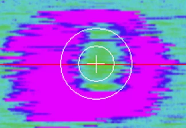
Image content: This image is available to view online.
View image online (https://assets.clevelandclinic.org/transform/a03d3405-275f-42a2-bc0f-17799190f860/19-EYE-4244-Talcott-CQD-Hero_jpg)
19-EYE-4244-Talcott-CQD-Hero
Researchers at Cleveland Clinic’s Cole Eye Institute are pioneering the use of novel image analysis systems to evaluate optical coherence tomography (OCT) for early detection of retinal disease and unique characterization of disease features.
Advertisement
Cleveland Clinic is a non-profit academic medical center. Advertising on our site helps support our mission. We do not endorse non-Cleveland Clinic products or services. Policy
Conventionally, OCT is used in clinical practice to visualize the retina, vitreoretinal interface and choroid. OCT provides outstanding visualization of retinal anatomy and is a critical piece to the evaluation of most retinal diseases.
Now, a new Cole Eye Institute study suggests that ellipsoid zone mapping, a new image visualization/analysis platform on OCT, may be an effective screening tool to identify subtle retinal changes indicative of toxicity from hydroxychloroquine treatment. The research was presented at the American Academy of Ophthalmology’s (AAO) 2019 annual meeting by Katherine E. Talcott, MD.
Hydroxychloroquine is commonly used to treat autoimmune inflammatory diseases such as rheumatoid arthritis. A rare side effect is retinal toxicity, which can cause irreversible and progressive vision loss. Detecting early signs of toxicity, before patients lose their vision, is critical to avoiding more severe vision complications. However, the early changes on OCT can be subtle and difficult to detect.
Cleveland Clinic Cole Eye Institute retina specialists Justis P. Ehlers, MD, who holds the Norman C. and Donna L. Harbert Endowed Chair for Ophthalmic Research, and Sunil K. Srivastava, MD, have developed an OCT analysis platform that integrates ellipsoid zone mapping technology that can be applied to OCT images to quantify metrics of various layers of the retina to better understand changes in disease.
Dr. Ehlers and his team have used the technology to detect alterations in the ellipsoid zone and outer retina in patients known to have hydroxychloroquine toxicity, but it has not been used to examine patients on hydroxychloroquine without known toxicity.
Advertisement
In the image analysis study described at AAO, a team led by Dr. Ehlers and Katherine E. Talcott, MD, through the Tony and Leona Campane Center for Excellence in Image-Guided Surgery and Advanced Imaging Research, examined the OCT scans of 401 patients who had been treated with hydroxychloroquine over a seven-year period and who also had OCT scans performed at different time points. The researchers applied the ellipsoid zone mapping software to the patients’ OCT images to determine if detectable longitudinal changes in the outer retinal bands were present.
The study’s goal was to assess if subclinical changes in ellipsoid zone integrity occurred and if this software tool was helpful in identifying these changes. “We actually were surprised that we were able to identify, in select patients, clear quantitative changes over time, even when the physician taking care of the patient didn’t recognize signs of toxicity,” Dr. Talcott says. “We found that this tool was able to pick up those changes.”
When evaluating the group overall, the researchers detected a significant increase in en face EZ attenuation from the first to the second OCT scan that correlated with the hydroxychloroquine-treated patients’ age (p = 0.01), daily dose (p = 0.01) and adjusted body-weight dose (p = 0.04). These findings suggest a progressive loss over time of EZ integrity. Of note, the features identified that correlate with increased attenuation are all risk factors for hydroxychloroquine toxicity.
Advertisement
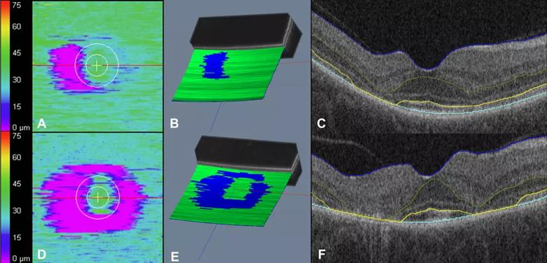
Image content: This image is available to view online.
View image online (https://assets.clevelandclinic.org/transform/9d8799af-cfce-48b2-9c02-f2304df9e7a5/19-EYE-4244-Talcott-CQD-Inset_jpg)
A case example of a 52 year old patient with hydroxychloroquine toxicity as seen on en face ellipsoid zone mapping. At baseline, the patient had evidence of ellipsoid zone attenuation (pink areas) seen on en face ellipsoid zone mapping (A), three-dimensional macular cube reconstruction (B), and on OCT images (C). The patient continued on hydroxychloroquine for 26 months after baseline. At follow-up, the patient had progressive ellipsoid zone attenuation (pink areas) seen on en face ellipsoid zone mapping (D), three-dimensional macular cube reconstruction (E), and on OCT images (F) signifying progressive toxicity. Source: Quantitative assessment of outer retinal layers and ellipsoid zone mapping in hydroxychloroquine retinopathy. Ugwuegbu O, Uchida A, Singh RP, et al. Br J Ophthalmol. 2019 Jan; 103(1):3-7. Used with permission.
Further research is needed before OCT can be applied as a screening tool. Next, Drs. Ehlers and Talcott plan to perform an in-depth analysis of this study cohort to evaluate whether these changes are amplified in specific patients or whether they represent global changes across the study population. In addition, they plan to evaluate whether specific values or quantitative findings can be used as cut-off values for subclinical toxicity. They also plan to evaluate the discordance between routine clinician evaluation of OCT grading and automated software-based assessment.
The goal is to better determine how to use this technology as a screening tool. “We need to hone in on what would be our cutoff for toxicity screening, and how we would better examine those patients who had had a change,” Dr. Talcott says.
Advertisement
“This tool could represent an important asset for clinicians to use in the screening for outer retinal diseases and retinal toxicity,” Dr. Ehlers says. “This could potentially be used to augment identification for patients at risk and/or patients with early toxicity. Optimized identification of those patients could reduce their risk of progression and additional visual loss.”
In addition to identifying OCT as a potential method for identifying hydroxychloroquine toxicity, the current study has other clinical implications, Dr. Talcott notes.
“More patients are probably having changes from this medication than we would expect,” she says. The researchers found that the mean dose being taken by patients in the study was only slightly less than the recommended dose. That means that nearly half of patients were taking more hydroxychloroquine than recommended. “The number of patients at risk could be higher than we anticipated,” she says. “We need better tools to screen for these patients.”
Dr. Ehlers can be reached at ehlersj@ccf.org and Dr. Talcott at talcotk@ccf.org.
Advertisement
Advertisement

The drug does not provide lasting disease control for most patients

New service for patients with diabetes will make annual screening more convenient

Gonioscopy can help determine next steps in reducing IOP

Similarly, patients with noninfectious uveitis have a higher risk of being diagnosed with an autoimmune disease within five years

A review of pathophysiology, risk factors, clinical presentation and treatment options

How computational tools and personalized biomechanics can improve keratoconus detection, ectasia risk assessment and surgical outcomes

Motion-tracking Brillouin microscopy pinpoints corneal weakness in the anterior stroma

Registry data highlight visual gains in patients with legal blindness