Locations:

Trauma patients present with infected pilon and distal femur fractures
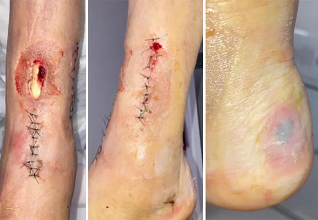
Image content: This image is available to view online.
View image online (https://assets.clevelandclinic.org/transform/87ed6cf0-41df-410f-940e-19d1bcecb4dd/23-ORI-4156369-CQD-650X450_jpg)
23-ORI-4156369 CQD 650X450
By Ronald Peirish, DO; Damien Billow, MD; Cesar Cereijo, DO; and Anokha Padubidri, MD
Advertisement
Cleveland Clinic is a non-profit academic medical center. Advertising on our site helps support our mission. We do not endorse non-Cleveland Clinic products or services. Policy
When treating patients with a nonunion, the clinical question is whether it is best to perform a primary amputation or arthroplasty, or to proceed with limb salvage. Limb salvage can involve treatment in a single- or two-stage procedure. Treatment is personalized, based on the patient’s unique injury characteristics and shared decision-making between the surgeon and patient.
Here we present three cases in which the decision was made to proceed with limb salvage.
A 35-year-old female presented to our emergency department for severe left ankle pain after sustaining a left closed pilon fracture four weeks earlier. She initially had been treated at an outside hospital with external fixation followed by open reduction and internal fixation (ORIF) four days later. Her postoperative course was complicated by wound dehiscence with positive cultures for Enterobacter, which was treated with vacuum-assisted closure (wound vac) and intravenous antibiotics.
Radiographs of her ankle demonstrated malreduction and fracture gapping of the articular surface (Figure 1). Physical exam revealed a 4.5 cm anterior pretibial wound with serosanguinous drainage and exposed anterior tibial tendon and hardware (Figure 2). Additionally, the patient had a stage 1/2 pressure wound to her heel.
Reconstruction commenced with irrigation and debridement, removal of hardware, and application of a uniplanar external fixator with wound vac. She underwent serial debridements. She then had a repeat ORIF (Figure 3) and synthetic bone graft with gentamycin once the wound bed was clean, followed by an anterolateral thigh (ALT) free flap by plastic surgery to cover the pretibial wound.
Advertisement
Postoperatively, her fracture and free flap healed with no concern for continued infection.

Image content: This image is available to view online.
View image online (https://assets.clevelandclinic.org/transform/9d271721-2872-4686-b29f-35eaf532b7f6/23-ORI-4156369-CQD-Inset1_jpg)
Figure 1. Radiographs demonstrate prior ORIF to the distal tibia with malreduction. Distal tibia intra-articular fracture demonstrates diastasis with medial column collapse and varus alignment of the ankle. Fibula demonstrates prior ORIF with apex posterior malreduction.

Image content: This image is available to view online.
View image online (https://assets.clevelandclinic.org/transform/fb8f366a-7720-4e29-b4b9-5cf0ecb66858/23-ORI-4156369-CQD-Inset2_jpg)
Figure 2. Clinical examination of the left ankle upon initial presentation to the emergency department. There is a 4.5 cm anterior pretibial wound with exposed anterior tibial tendon (A). Previous nylon suture is in place (B). There is a stage 1/2 pressure wound to the heel (C).

Image content: This image is available to view online.
View image online (https://assets.clevelandclinic.org/transform/33d525a6-c724-4450-9759-0abebdcbc208/23-ORI-4156369-CQD-Inset3_jpg)
Figure 3. Three-month postoperative radiographs demonstrate callus, neutral alignment and intact hardware to the tibia. Vascular clips present.
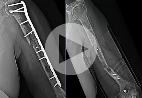
A 70-year-old female with a medical history of smoking, deep vein thrombosis, chronic obstructive pulmonary disease, stage 3 chronic kidney disease and rheumatoid arthritis presented to our clinic for evaluation of continued pain and ambulatory dysfunction of her left lower extremity. After a left distal femur fracture, she had been treated at an outside hospital with a distal femoral locking plate. Her postoperative course was complicated by nonunion with hardware failure (Figure 4). She was treated with a bone stimulator and knee brace.
In our office, the patient stated that she could ambulate short distances with a walker but mostly used a wheelchair. On exam, her surgical incisions were well healed. The range of motion of her knee was 0-90 degrees of flexion with gross motion at the fracture site. Surgical intervention was recommended as the fracture was unlikely to heal due to instability at the fracture site.
The patient was taken to the operating room, where the previous hardware was removed, the nonunion site was debrided, the fracture was reduced, and a retrograde femoral nail was placed. Bone graft was applied to the fracture site from the left femur using a reamer-irrigator-aspirator (RIA) system.
Postoperatively, the fracture healed with maintained alignment (Figure 5). At her last clinic appointment, the patient was pain-free and ambulating with a walker.

Image content: This image is available to view online.
View image online (https://assets.clevelandclinic.org/transform/771008ed-2929-4722-99a7-0cb6f5b8dc57/23-ORI-4156369-CQD-Inset4_jpg)
Figure 4. Preoperative radiographs of the left distal femur demonstrate a supracondylar nonunion with flexion and valgus alignment. There is failure of a lateral distal femur locking plate with multiple broken screws proximal to the fracture site. Chondrocalcinosis of the knee with joint space narrowing is evident.

Image content: This image is available to view online.
View image online (https://assets.clevelandclinic.org/transform/5447b676-f0c9-4b62-a044-a93db2031983/23-ORI-4156369-CQD-Inset5-766x1024_jpg)
Figure 5. Postoperative radiographs of the left distal femur demonstrate abundant callus of the left distal femur fracture with near anatomic alignment. Hardware is intact, without evidence of loosening or breakage.
A 32-year-old female presented to our office with a history of polytrauma. She had been hit by a motor vehicle and had sustained a right open distal femur fracture (grade 3A), right patella fracture, left closed midshaft femur fracture and bilateral distal radius fractures. Her right femur was treated at an outside hospital with a retrograde femoral nail and a two-stage Masquelet procedure to address a large bone void.
Advertisement
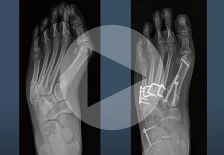
The patient presented to our office for continued right distal thigh pain, ambulatory dysfunction and a limb length discrepancy of 3 cm (Figure 6). Radiographs demonstrated an 8 cm bone void with nonunion (Figure 7).
Preoperative surgical planning commenced, and it was decided that a custom synthetic graft cage would be created to add 2 cm of length to her right lower extremity.
The patient was taken to the operating room, where the previous retrograde nail was removed, an osteotomy was created, and the femur was lengthened acutely using a femoral distractor. Bone graft was harvested from the femur and ipsilateral tibia by RIA. A new retrograde nail with a side plate was implanted into the femur. The bone graft was mixed with synthetic bone graft and impacted into the custom cage, which was inserted into the femur and secured with screws.
Postoperatively, the patient had two distal interlocking screws that loosened and became prominent. As a result, she had a subsequent surgery to remove the prominent screws. The patient progressed to full weight-bearing, and her last radiographs demonstrated maintained alignment with incorporation of the bone graft with bone bridging (Figure 8).

Image content: This image is available to view online.
View image online (https://assets.clevelandclinic.org/transform/56e52404-8df3-4b4a-87d7-62a10e76857a/23-ORI-4156369-CQD-Inset6-450x1024_jpg)
Figure 6. Radiograph of the lower extremities demonstrates bilateral retrograde femoral nails. Right femur demonstrates 3 cm shortening compared to the left femur, with 8 cm bone void. Hardware is bilaterally intact without loosening. Left midshaft femur demonstrates callus and bone bridging.

Image content: This image is available to view online.
View image online (https://assets.clevelandclinic.org/transform/7e936608-2bb6-4212-ac4a-69d653bfff64/23-ORI-4156369-CQD-Inset7_jpg)
Figure 7. Preoperative radiographs of the right femur demonstrate retrograde femoral nail with 8 cm bone void and intact hardware to the patella.

Image content: This image is available to view online.
View image online (https://assets.clevelandclinic.org/transform/36710485-d546-46a9-ba7f-44e591d7b038/23-ORI-4156369-CQD-Inset8_jpg)
Figure 8. Postoperative radiographs of the right femur demonstrate maintained alignment with retrograde femoral nail and distal femoral side plate. Bone graft is present with bone bridging.
These three cases demonstrate that nonunions can cause severe dysfunction and pain.
Limb salvage is a viable option for certain patients, helping them return to function by preserving their native extremity.
Drs. Billow, Cereijo and Padubidri are orthopaedic traumatologists at Cleveland Clinic. Dr. Peirish is a trainee in Cleveland Clinic’s orthopaedic residency program.
Advertisement
Advertisement

Relieves discomfort, reduces opioid dependency and improves quality of life

A hand and wrist surgeon explains different approaches based on nature and severity of injury
Finally, a solution after multiple revision surgeries for delayed bone healing, loose hardware and unrelenting infection
Surgeon corrects skew foot to address repeat injuries

Innovative procedure offers less-invasive alternative to Latarjet procedure

Even patients with reported penicillin allergies can receive it without increased complications

Study challenges assumptions about risk evaluation in total hip revision

Protein expression in synovial fluid indicates patients’ immune factors may be involved