Locations:

Multicenter collaboration aims to facilitate tracking of neurological activity deep within tissue
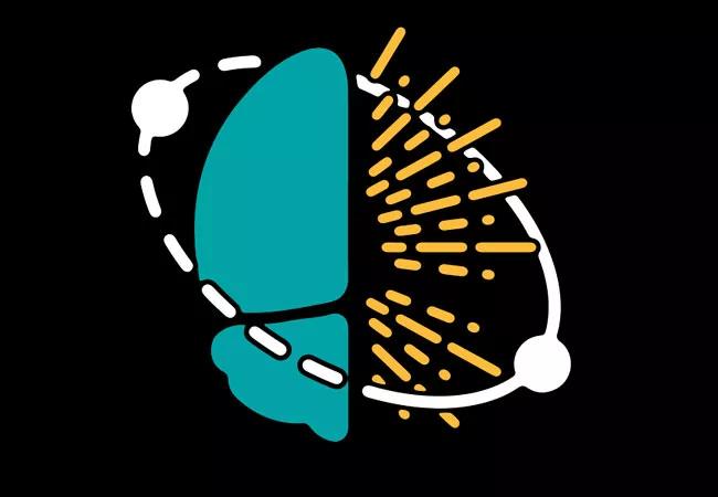
Image content: This image is available to view online.
View image online (https://assets.clevelandclinic.org/transform/fa36b7fb-a50a-4ea5-8ee3-89907e326f2d/23-NEU-4202909-CQD-Hero-650x450-1_jpg)
23-NEU-4202909-CQD-Hero-650×450
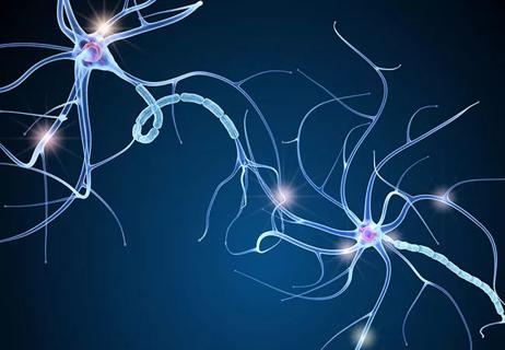
To better understand neurological disorders through microscopy, scientists need to be able to capture brain activity in a matter of milliseconds and deep within live tissue. An ongoing research collaboration between Cleveland Clinic, Harvard University and the Massachusetts Institute of Technology (MIT) is developing microscopes and artificial intelligence (AI) models that can rise to the task.
Advertisement
Cleveland Clinic is a non-profit academic medical center. Advertising on our site helps support our mission. We do not endorse non-Cleveland Clinic products or services. Policy
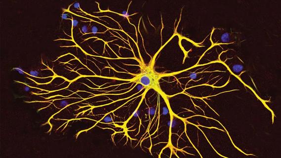
Their approach, called “Deep Learning Powered De-scattering with Excitation Patterning” (DEEP2), uses ultrafast lasers to penetrate light throughout a tissue specimen at larger scale, taking thousands of measurements. These measurements are then rapidly translated to images using a deep learning algorithm trained on large data sets.
The researchers recently unveiled DEEP2 in the journal Light: Science & Applications (2023;12:228). DEEP 2 is an update to a model named “De-scattering with Excitation Patterning” (DEEP) that the team published two years ago (Sci Adv. 2021 Jul 7:7[28]:eaay5496). DEEP’s updated algorithm produces clearer images based on fewer data points.
“Current techniques can’t reach the depth and speed we need to advance our understanding of neurological conditions such as autism spectrum disorders, Alzheimer’s disease and multiple sclerosis,” says co-investigator Murat Yildirim, PhD, of the Department of Neurosciences in Cleveland Clinic Lerner Research Institute. “By creating and refining microscopy techniques integrated with computational imaging techniques like DEEP2, we are providing invaluable clinical context for patient care.”
Dr. Yildirim’s team spearheaded the DEEP microscope’s design and its use in preclinical models and worked with colleagues at Harvard and MIT to train and develop the accompanying computational model.
The DEEP approach was developed to overcome limitations of point-scanning multiphoton microscopy, which has been the gold standard for imaging biological structures deep inside tissue. The latter technology focuses laser light at long wavelengths through scattering tissue to excite fluorescent molecules. The microscope then collects emission fluorescence through the same scattering tissue onto a detector. However, because the emission light is at much shorter wavelengths, it undergoes substantial scattering en route to the detector, which prevents excitation and resolution of multiple points at the same time. This requires sequential imaging, one point at a time, which makes point-scanning multiphoton microscopy a slow imaging technique. If speed is necessary, compromises must be made to either image resolution or the field of view.
Advertisement
In contrast, DEEP was developed as a wide-field microscopy alternative to point-scanning technology. DEEP uses patterned multiphoton excitation with long wavelengths to encode spatial data inside tissue before scattering. However, de-scattering at typical tissue depths requires hundreds of these patterned excitations.
To overcome this requirement, the researchers integrated DEEP’s wide-field multiphoton microscopy with a deep-learning algorithm to create DEEP2, which can reconstruct a de-scattered image from only 32 DEEP measurements, versus 256 measurements with the original DEEP model. “DEEP2 is nearly an order of magnitude faster than the original DEEP,” Dr. Yildirim observes.
In their newly published study, the researchers validated their approach using experimentally acquired test data with a custom-made three-photon microscope that Dr. Yildirim developed. They demonstrated that DEEP2 can de-scatter in vivo cortical vasculature images in mice up to four scattering lengths below the surface.
“Our work sets the foundation to develop data-driven inverse solvers for DEEP temporal focusing microscopy using deep learning,” the researchers write, “and hence is an important milestone for in-vivo wide-field multiphoton microscopy.”
Dr. Yildirim’s lab also works on other computational models at Cleveland Clinic as part of the Discovery Accelerator, a research partnership with IBM, to push the limits of deep tissue imaging.
“We can harness how quickly artificial intelligence and deep learning are advancing in this type of imaging,” Dr. Yildirim says. “These advanced technologies ― including quantum computing ― open a door for us to understand health problems in a way like never before. The tools are there; we just need to build and refine the best ways to use them.”
Advertisement
Advertisement

Phase 3 STELLAR trial underscores role of molecular stratification in glioma care

A principal investigator of the landmark longitudinal study shares interesting observations to date
GFAP elevation may signal increased risk of progressive regional atrophy, cognitive decline

Preclinical work promises large-scale data with minimal bias to inform development of clinical tests
Research aims to extend observations of reversal learning in mice to human neurological disorders
Diagnosis and treatment of MOG antibody-associated disease

Designed for digitization, distance health, discovery and more
Model of viral encephalomyelitis shows links with CNS recovery from inflammation and damage