Locations:





Cleveland Clinic findings highlight gains in stability, chest wall contouring and correction
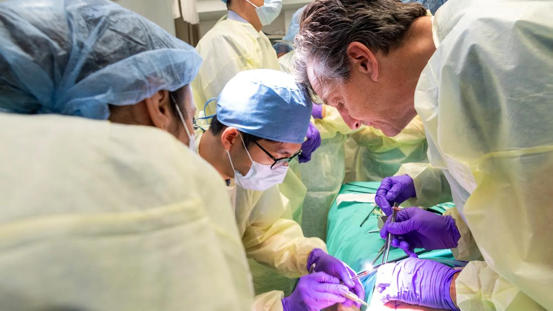
Image content: This image is available to view online.
View image online (https://assets.clevelandclinic.org/transform/dca0e68a-650a-4411-9b7a-2e298e7e09d0/CHP_5003858_06-28-24)
Surgeons in a training session
John DiFiore, MD, (right) Director of Cleveland Clinic's Pectus Excavatum Program, demonstrates the NeoPectus system during an instructional session in a simulation lab.
Advertisement
Cleveland Clinic is a non-profit academic medical center. Advertising on our site helps support our mission. We do not endorse non-Cleveland Clinic products or services. Policy
The NeoPectus method developed by South Korean surgeon Hyung Joo Park, MD, PhD, during the past decade has revolutionized minimally invasive pectus excavatum (PE) repair, virtually eliminating the problem of bar displacement and enabling more extensive, functional and aesthetic chest wall remodeling.
Despite those benefits, adoption of the NeoPectus approach beyond parts of Asia and Europe has been slow, likely due to lack of awareness and the technique’s learning curve.
After training with Dr. Park in South Korea in 2023, John DiFiore, MD, the Director of Cleveland Clinic’s Pectus Excavatum Program, brought the NeoPectus method to the United States. He has since performed more than 150 procedures, recently publishing the results of the first 72 cases.
Dr. Park’s system “is a paradigm shift” in PE correction, Dr DiFiore says. Cleveland Clinic has embraced it because “we embrace new ideas, new concepts, new techniques. That’s ingrained in our DNA.”
With Dr. Park’s recruitment to Cleveland Clinic in 2025 and the arrival of Manuel López, MD, the past president of the Chest Wall International Group, “between the three of us, we have 80 years of pectus experience,” Dr. DiFiore says. “We're operating together, innovating week to week and learning from each other. I think we have the most experienced pectus team in the world at this point.”
Drs. DiFiore and Park conduct biannual NeoPectus workshops for visiting surgeons. “One of my goals is for our pectus program to become a teaching center for the technique,” Dr. DiFiore says, “and through our research papers, to present our concepts on an international stage.”
Advertisement
PE, characterized by varying degrees of posterior sternal depression, is the most common congenital chest wall deformity. The condition has both physical and psychological consequences, including impaired breathing and cardiac function, fatigue, chest pain, poor self-esteem and depression.
Surgical efforts to correct PE date back more than a century.
The initial technique, introduced in 1911, involved resection of the second and third costal cartilages. It was later modified to include sternotomy and external sternal traction.
Pediatric surgeon Mark Ravitch’s approach, which became the standard of care for decades after publication of the first case series in 1949, called for surgical removal of all costal cartilages and a transverse wedge osteotomy of the sternum. This eliminated the need for sternal traction but did so with a challenging and very invasive surgical procedure.
Seeking a less radical and invasive means of PE repair, pediatric surgeon Donald Nuss in 1987 employed a convex titanium bar threaded through a tunnel he created under the sternum at its deepest depression point. Flipping the bar to a concave position elevated the sternum and corrected the PE without cartilage or rib resection or sternal osteotomy, and with minimal blood loss.
The Nuss procedure quickly was adopted worldwide and largely supplanted the Ravitch approach. Unfortunately, despite various methods meant to anchor the bar including fixators, lateral stabilizers and pericostal sutures, it would slip out of place in as much as 27% of cases. Bar displacement can cause intercostal muscle stripping, cardiac or aortic injury and other serious complications that necessitate reoperation.
Advertisement
Dr. Park aspired to create a minimally invasive PE repair strategy that not only solved the bar displacement problem but was able to produce a functionally and anatomically normal and aesthetically pleasing chest wall.
Ultimately, the NeoPectus system he developed involves four steps:
Advertisement
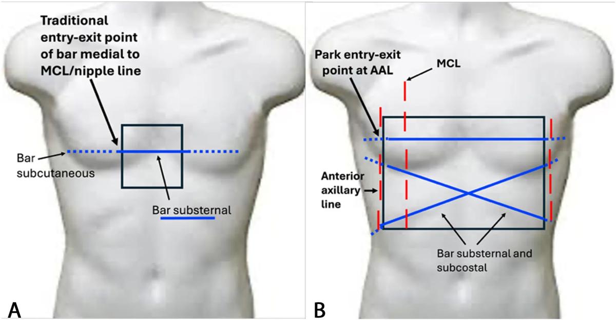
Image content: This image is available to view online.
View image online (https://assets.clevelandclinic.org/transform/391f8613-2b01-4d96-a4d3-4b914b080df3/26-CHP-7821058-park-procedure)
A. Traditional single bar with entry/exit point at or medial to the mid-clavicular line limits corrected area to the central sternum. B. NeoPectus system with three bars and entry/exit points in the region of the anterior axillary line significantly increases total area of chest wall correction. From: DiFiore J, Heitsman C, Robertson J Parks bridging bar pectus procedure: Techniques to optimize functional and aesthetic results in 72 cases; Journal of Pediatric Surgery, 2025; p. 61
Dr. Park described his technique at the 2021 Chest Wall International Group annual meeting.
“I heard that lecture and it was like a light bulb went off,” Dr. DiFiore recalls. “I said, ‘Well, this is far superior to what we're doing.’ In addition to bar displacement being eliminated by the use of bridging bars, the bridges also allows us to move the entry and exit points of the bars more laterally to correct a larger surface area of the chest. The benefits are functional and aesthetic. It increases the volume of a larger area of the chest wall to maximally improve lung expansion. And it allows a much broader approach to chest wall contouring — in particular, a way to treat rib flare or costal flaring that was not addressed with prior techniques."
Dr. DiFiore invited Dr. Park to Cleveland Clinic, where he gave a lecture and demonstrated the NeoPectus hardware. Subsequently, in January 2023, Dr. DiFiore spent two weeks with Dr. Park in South Korea, participating in multiple NeoPectus surgeries and learning more about his techniques.
When Dr. DiFiore returned home, he and his surgical team began performing PE repairs with the NeoPectus approach — its first use in the United States.
In his report of the initial 72 cases performed at Cleveland Clinic between January 2023 and May 2025, Dr. DiFiore noted that there were no instances of bar displacement, revision surgery or device-related complications during the study’s 2.4-year follow-up period. Most patients were discharged on the first postoperative day.
Advertisement
The lack of displacement was notable considering that 60% of patients in the study cohort required complex crossed-bar configurations. (There have been no displacements in patients who have undergone NeoPectus surgery since the study’s conclusion, Dr. DiFiore says.)
Pain management included preoperative dosages of gabapentin, acetaminophen and celecoxib; intraoperative thoracoscopic cryoablation of intercostal nerves and intercostal nerve blocks; and postoperative use of diazepam and oxycodone. Dr DiFiore also published this technique in 2022 showing that with this cryoablation protocol, patients can be discharged home the day after surgery.
The bridge plates that anchor the pectus bars in the NeoPectus procedure don’t increase pain levels beyond that of traditional pectus surgery, Dr. DiFiore says. However, initial patients in the study experienced heightened pain in the lower chest wall in the area where compression sutures were used to smooth costal flares. To compensate, Dr. DiFiore extended cryoablation distally from the eighth to the 10th rib, which provided additional relief.
Pectus bars typically remain in place for three years. Some surgeons have questioned whether costal flaring will return when the compression sutures are removed. “Anecdotally, I’ve now removed the bars in a handful of patients and have not noticed a recurrence of rib flaring,” Dr. DiFiore says. “But that is one of the things we will study going forward. We measure the angle of the protruding rib before and after surgery and when the bars are removed to see if there's recurrence. We're actually putting numbers to it.”
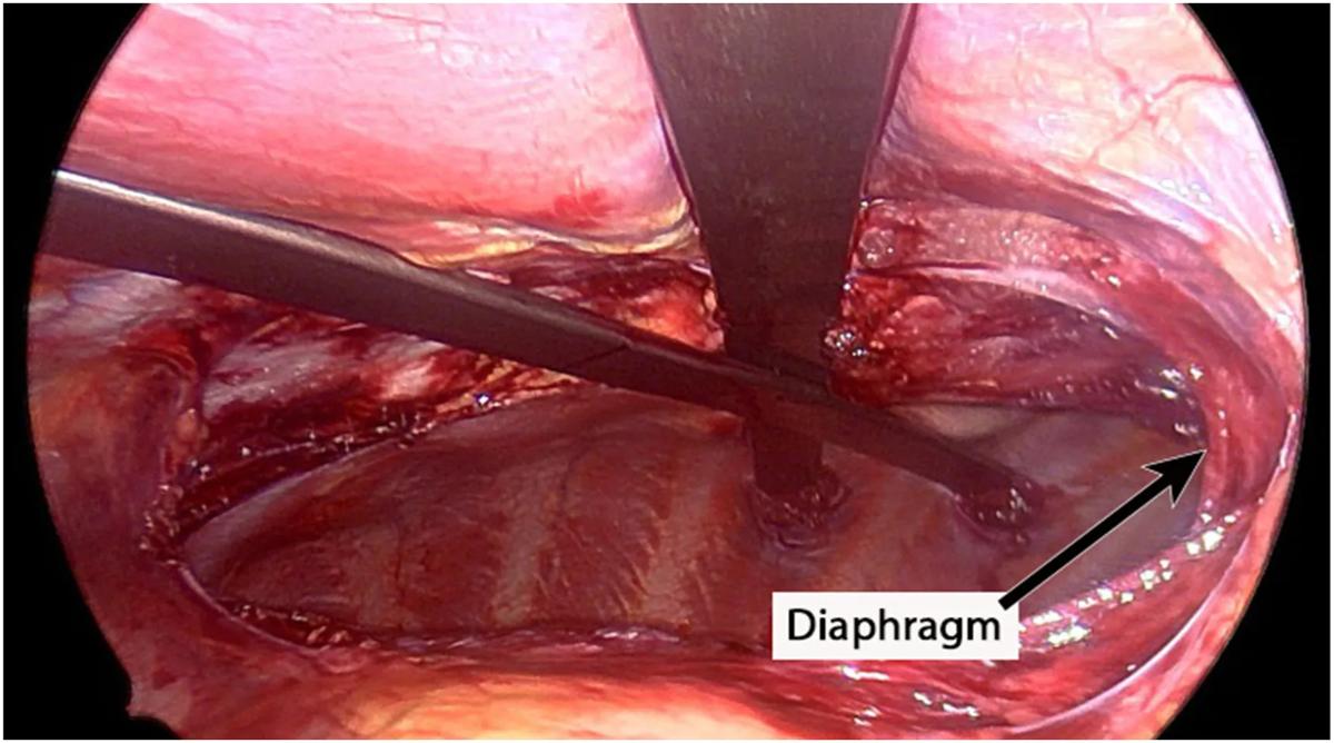
Image content: This image is available to view online.
View image online (https://assets.clevelandclinic.org/transform/a7347b21-11c4-4853-9634-daf2186af731/26-CHP-7821058-park-procedure-scope)
Completed dissection with crossbars in position. Arrow points to insertion of diaphragmatic muscle fibers onto the posterior sternum. From: DiFiore J, Heitsman C, Robertson J Parks bridging bar pectus procedure: Techniques to optimize functional and aesthetic results in 72 cases; Journal of Pediatric Surgery, 2025; p. 61
The traditional approach to PE repair has sometimes meant a tradeoff between functional improvement and cosmetic gains.
“When you have a very inferior or low point of maximum depression, that’s where you get maximal compression of the heart,” Dr. DiFiore explains. Before the advent of the NeoPectus method, “there were multiple cases where we would put a bar across that low point and get nice elevation, but the costal margin would be overextended to a point that was aesthetically unacceptable. We would then have to remove the bar and move it back a space so it was aesthetically at least no worse than when we started. In a lot of situations that led to inadequate physiologic decompression of the heart.”
Now, though, “we can place a bar wherever we want,” he says. “If the bar is right on the costal margin and the ribs come out even further, we can fix that. It frees us up to create a more physiologically sound operation with no compromises in our cardiac decompression. We can keep the bar right where it needs to be and deal with the associated rib protrusion without any problems.
“When I lecture about this, I advise surgeons to attack the point of maximal depression with their dissection and bar placement, understanding that no matter where you put that bar, if you put another bar and bridge them together, it's going to be stable. So, basically, your bar positioning is not limited by anything.”
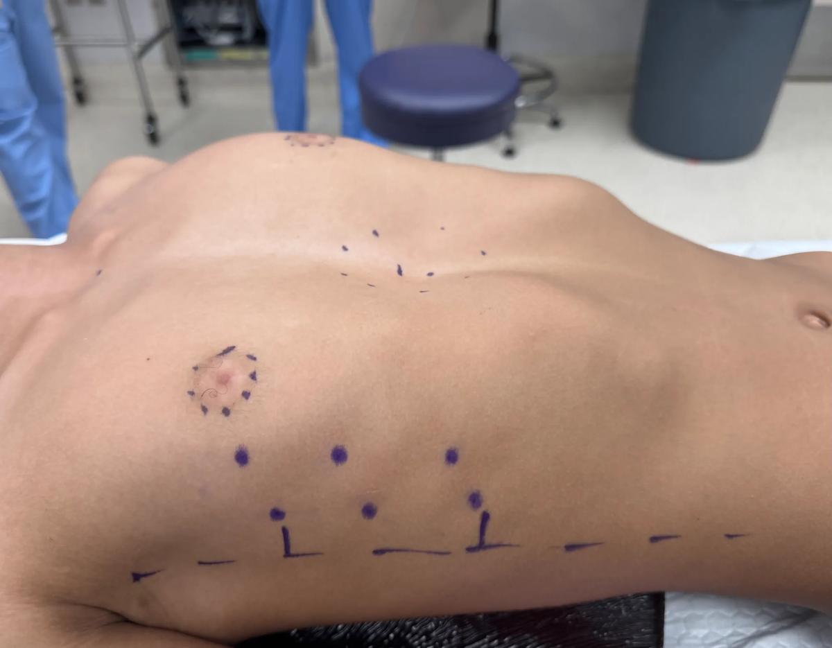
Image content: This image is available to view online.
View image online (https://assets.clevelandclinic.org/transform/cb7bf739-f462-43bf-ace0-f0ecd9e06256/chest-pre-4518)
Chest wall prior to repair shows costal flaring.
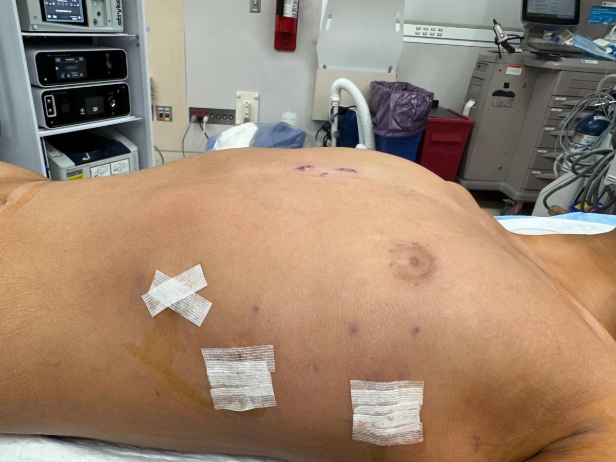
Image content: This image is available to view online.
View image online (https://assets.clevelandclinic.org/transform/24106aaf-8cb3-4c73-9b20-c5e46fa29b1c/chest-post-4520)
Chest wall after NeoPectus repair shows rib flaring resolved.
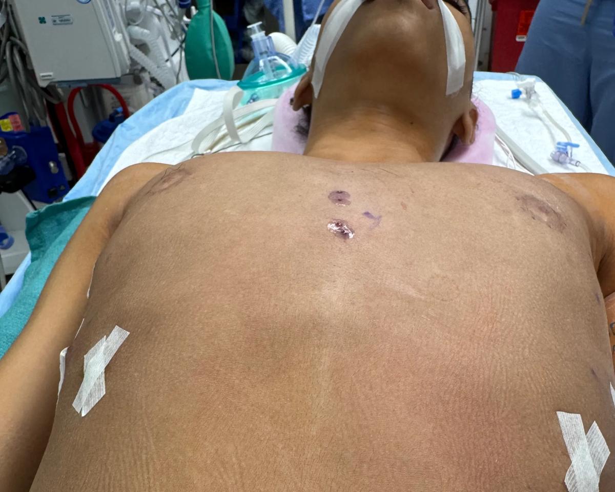
Image content: This image is available to view online.
View image online (https://assets.clevelandclinic.org/transform/f8f4bf01-8717-4e33-99bf-cd2f1ba25ec3/chest-post-4522)
Frontal view showing chest wall after NeoPectus repair.
The versatility of the NeoPectus approach has increased surgeons’ comfort with talking to patients about the aesthetic potential of PE repair.
“For the longest time, ‘aesthetic’ was considered a dirty word in pectus surgery,” Dr. DiFiore says. “It was like, ‘Well, we don't care about that; we just want physiological improvement.’ But that’s shortsighted. Patients should want their chest to look normal as well as to be functionally sound, and now we can provide both.
“One of my personal goals in educating the pectus community is that aesthetics are just as important as the functional results,” he says. “I used to tell patients, ‘Don’t expect a completely normal chest — that's unrealistic.’ In the last three years with the introduction of Dr. Park’s system, we are coming as close to a perfect repair as we can get.”
Advertisement
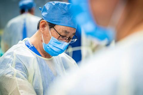
Cleveland Clinic welcomes world-leading chest wall surgeon Dr. Hyung Joo Park

What the discovery means for clinicians and patients

Why preparedness matters right now

Early diagnosis, specialized assessment and high-frequency motor-based therapy can improve outcomes

Transplant centers vary substantially in how they assess, document and follow patients after allogeneic transplantation

Helping pediatricians and families make safer, more individualized activity decisions

Study finds exome sequencing can uncover additional diagnoses after nondiagnostic panel testing

Early planning can help prevent disruptions in treatment, medication access and support
 Search