Locations:

A physician's perspective on confirmation imaging, diagnostic confidence and choosing the right robotic platform for patients
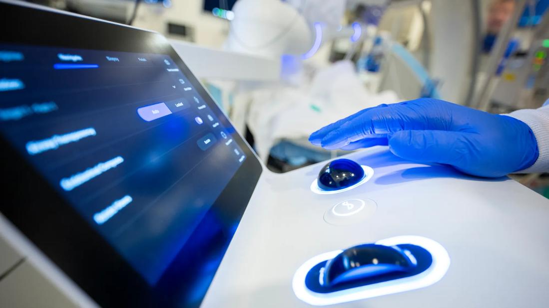
Image content: This image is available to view online.
View image online (https://assets.clevelandclinic.org/transform/e090724c-181d-4ba9-a93c-6cb87101488c/PUL_5529311_02-18-25_0009_MK)
Controls for a robotic-assisted bronchoscopy platform
Written by See-Wei Low, MD, and Colin Gillespie, MD
Advertisement
Cleveland Clinic is a non-profit academic medical center. Advertising on our site helps support our mission. We do not endorse non-Cleveland Clinic products or services. Policy
We have been fortunate to gain hands-on experience with the three robotic-assisted bronchoscopy (RAB) platforms currently used in clinical practice: the electromagnetic navigation–based Monarch system (Johnson & Johnson/Auris) and Galaxy platform (Noah Medical), and the shape-sensing Ion platform (Intuitive Surgical). From a technical standpoint, all three can be used successfully to navigate to peripheral pulmonary lesions (PPLs).
As RAB has rapidly matured—moving from novelty to routine practice—it has become clear that navigation alone is not the finish line. For patients and providers, the real question is not whether we can localize a lesion, but whether we can consistently obtain adequate tissue for diagnosis and ancillary testing while doing so safely.
That distinction becomes obvious when reading—and trying to apply—the growing RAB literature. Diagnostic yield across studies often appears reassuringly similar, and major complications such as pneumothorax or clinically significant bleeding are uncommon. Yet these headline numbers rarely tell the whole story.
Yield is not a fixed property of a platform; it reflects a complex interaction among lesion characteristics, patient factors, operator experience, imaging strategy and institutional workflow.
Lesion size and location, proximity to the pleura, and the presence of a bronchus sign all influence the technical difficulty of reaching and engaging the target. Patient-level factors such as emphysema, prior radiation, anticoagulation and obesity can affect both feasibility and procedural risk. Just as importantly, operator judgment—knowing when to persist, reposition, change strategy, or stop—often determines whether a procedure ends in success or a missed opportunity.
Advertisement
Adjunctive imaging adds another layer of complexity. Radial EBUS (r-EBUS), fluoroscopy, cone-beam CT (CBCT) and tomosynthesis-style confirmation each provide different perspectives on localization. But each comes with its own tradeoffs in time, cost, radiation exposure, staffing and logistics. Studies also vary in how they define “diagnostic yield” and how rigorously negative results are verified. When definitions, follow-up and case mix differ, platform-to-platform comparisons become difficult to interpret.
In day-to-day practice, it becomes apparent that “platform performance” is tightly intertwined with imaging and three-dimensional localization. In many ways, overall program performance depends on the successful alignment of imaging, sampling technique, pathology workflows and multidisciplinary coordination.
Seen through this lens, robotic platforms feel less like direct competitors and more like different routes to the same goal: a consistent, efficient diagnosis with adequate tissue acquisition delivered with an acceptable safety profile.
The practical question becomes not which robot is “best,” but which system integrates most naturally into a given program and reliably converts navigation into meaningful clinical information.
In Dr. Low’s experience, Ion can feel particularly intuitive early on, and many operators appreciate its workflow and shape-sensing approach. Its non-electromagnetic design is also attractive in day-to-day practice, and some users find the learning curve for distal navigation manageable. When paired with strong intraprocedural confirmation imaging, Ion can support a reproducible sequence that moves logically from navigation to confirmation to sampling. In programs with consistent access to CBCT (mobile or fixed) and teams already comfortable integrating that imaging, the overall process can feel structured and controlled—while recognizing that diagnostic success depends on multiple factors beyond the platform itself.
Advertisement
That said, platform choice is rarely one-size-fits-all. Not every center has reliable access to CBCT or the staffing and throughput to integrate it seamlessly into routine cases. In those settings, the practical advantages of alternative workflows become more prominent. Galaxy is particularly interesting because it emphasizes lesion confirmation using existing two-dimensional fluoroscopy, aiming to narrow the gap between navigation and true lesion engagement without major infrastructure demands. While early experience is encouraging, broader adoption should be guided by larger independent multicenter datasets using standardized definitions and clinically meaningful verification of lesion engagement and sampling adequacy.
Monarch benefits from the longest real-world experience and operational familiarity at many institutions, which can translate into efficiency and consistency—especially in complex or high-volume programs. Monarch Quest has also drawn interest because it is designed to expand imaging integration, including the ability to incorporate three-dimensional imaging into the procedural workflow, which may strengthen confirmation strategies in centers that can support it. We cannot verify comparative performance claims for Quest across institutions, but conceptually, the direction aligns with where the field is heading: tighter coupling of navigation with robust, repeatable confirmation. As with any platform, outcomes ultimately depend less on the robot itself than on how consistently confirmation and sampling are pursued, along with team experience, imaging access, and case selection.
Advertisement
What has become increasingly clear is how profoundly confirmation imaging changes behavior. The difference between “we navigated to the target” and “we confirmed tool-in-lesion in three planes” is not subtle. The ability to demonstrate localization and tool-in-lesion influences decisions at every step of the procedure and shapes how we counsel patients afterward. Confirmation is not just an adjunct anymore - it is central to diagnostic confidence - regardless of platform.
Looking ahead, the field would benefit from greater standardization. We need harmonized definitions of diagnostic yield, consistent reporting of tool-in-lesion confirmation, transparent reporting of lesion and patient complexity, and adequate follow-up to adjudicate false-negative results. Pragmatic multicenter registries and comparative studies - including randomized trials that account for imaging strategy, sampling tools, pathology workflows, radiation exposure and procedural cost (in addition to yield) will be essential to optimize our role within thoracic oncology.
As bronchoscopic ablative therapies for PPLs continue to evolve, the importance of stable navigation and reliable confirmation will only grow. Ultimately, the future of RAB lies less in platform superiority and more in how consistently a program can translate navigation into diagnosis and potentially therapy - while keeping patient risk central to every decision.
Advertisement
Advertisement

Appropriate patient selection and clinician awareness remain key to broader use
Caregivers are provided with real-time bronchoscopy patient findings

The role of interventional pulmonology in lung nodules/lesions care

Common misconceptions about perioperative management and strategies to improve care

Dr. Prabhu discusses mentorship, collaboration and her vision for the future of the department

Multidisciplinary collaboration is fueling breakthroughs in endoscopic and surgical technology

Study finds broadly similar outcomes between MIS and open surgical approaches

Join us in Cleveland July 17 for a practical, first-of-kind course