Locations:





Two cases show how integrated surgical and prosthodontic planning is necessary for dental rehabilitation of major maxillary defects
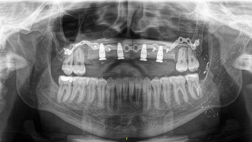
Image content: This image is available to view online.
View image online (https://assets.clevelandclinic.org/transform/adb4192c-9baa-4e4a-9a78-5020c2e65a56/26-HNI-8330108-ReconstructionDeficientMaxilla-Tso-feature_967x544)
Panoramic radiograph showing the completed midface reconstruction
Complex maxillofacial reconstruction requires careful attention to both function and appearance. Patients with major palatal or maxillary defects may live without teeth or with compromised speech, chewing, and breathing, particularly after cancer surgery and radiation therapy. Acquired facial deformity affects quality of life. For these reasons, coordinated planning among head and neck surgery, facial plastics and dental teams is essential to support functional rehabilitation and an aesthetic outcome.
Advertisement
Cleveland Clinic is a non-profit academic medical center. Advertising on our site helps support our mission. We do not endorse non-Cleveland Clinic products or services. Policy
Two recent Cleveland Clinic cases show how early reconstructive and prosthodontic planning can expand what is possible for patients with complex defects.
A 29-year-old patient presented to Cleveland Clinic’s Department of Facial Plastic Surgery for management of midface/maxillary deficiency in 2021. The patient’s main goals were correction of his midface deformity and to have teeth that stayed in.
“The patient was born with a primary palate teratoma that was resected when he was 3 months old,” explains Theodore Tso, DMD, the patient’s treating maxillofacial prosthodontist. “Since then, he had a significant midfacial defect and lack of maxillary anterior teeth. The patient was from out of state but came to us in 2021 after coming across an article involving fibula flap transfers from my colleague, Michael Fritz, MD, FACS, the Director of the Section of Facial Plastic and Microvascular Surgery at Cleveland Clinic.”
The patient had treatment with a local orthodontist to level and align his teeth before coming to see Dr. Tso in 2024.
“For a young patient with high aesthetic expectations, every part of the surgery had to be planned with a high degree of precision,” explains Dr. Tso. “The placement of the bone, soft tissue flap and dental implants all had to support optimal front tooth appearance and facial contours.”
Because the patient had specific goals for both his teeth and facial appearance, the team first made a partial denture to establish the desired tooth position and upper lip projection. This partial denture was then scanned and merged with bone data from a cone-beam computed tomography scan. They then used a fully guided CT-based approach to simulate the surgery on models before entering the operating room.
Advertisement
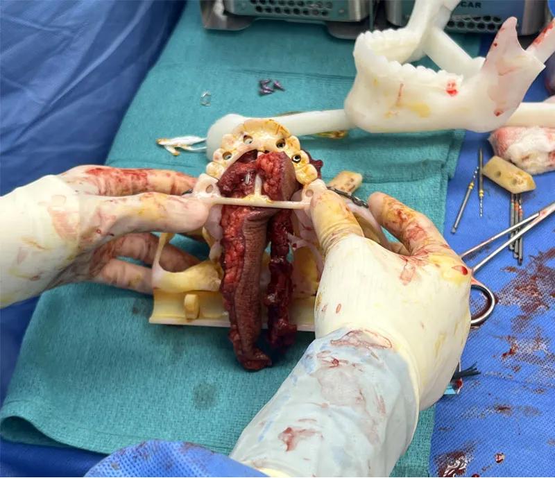
Image content: This image is available to view online.
View image online (https://assets.clevelandclinic.org/transform/95358c4d-2d4e-4581-b992-f9bffb98a8ee/ReconstructionDeficientMaxilla-Tso-CQD-Inset1_800x689)
The fibula flap with dental implants is assembled with a 3D printed model in the operating room to confirm the positioning aligns with what was planned.
The planning sequence worked backward from the desired final tooth position. Once the team knew where the teeth needed to be, they could determine where to place the implants and the underlying bone brought up from the leg.
“Tooth position dictates where the implants go, and that dictates where the fibula flap is placed,” he explains. “In these cases, it can be difficult to predict the projection and volume of the lip and soft tissue. From the microvascular surgeon’s side, it is also technically challenging to place a leg bone into the front of the upper jaw, so this case required extensive preoperative planning.”
In late 2024, the patient underwent surgery involving teeth extraction, infrastructure maxillectomy, vestibuloplasty, transfer of a fibula flap, and placement of dental implants. The surgery was a three-team procedure — the resective piece was done by Sara Liu, MD, a facial plastic and reconstructive surgeon; the microvascular reconstruction piece was done by Dr. Fritz; and the prosthodontics piece was done by Dr. Tso.
By mid-2025, the patient’s teeth had been completed. Dr. Tso reports that the patient is now able to chew and speak normally. He has a good dental appearance and midface contour.
“One of the biggest challenges in this case was the aesthetic component,” explains Dr. Tso. “We see referrals for patients who have had similar surgeries elsewhere where the midface may appear too projected or too collapsed. Avoiding that was a major goal. That is where multidisciplinary collaboration is especially valuable — it helps ensure that functional and aesthetic goals are addressed together.”
Advertisement
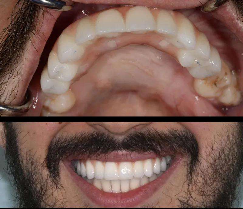
Image content: This image is available to view online.
View image online (https://assets.clevelandclinic.org/transform/1dc4c1be-8f9d-47dc-aebd-d4f4af42a786/ReconstructionDeficientMaxilla-Tso-CQD-Inset3_800x689)
Above: Intraoral view of the patient's completed treatment. Note the dental and palatal anatomy closely approximate normal contours. Below: Successful treatment gives the patient good dental esthetics as well as midface projection
“This case showcases our ability to achieve reconstructive outcomes that optimize both function and aesthetics without compromising on either,” adds Dr. Liu. “That's only been possible with close collaboration between facial plastic and reconstructive surgery and prosthodontics.”
A 78-year-old patient presented to Cleveland Clinic Head and Neck Surgery in 2024 with a large exophytic growth at the anterior palate. The growth led to a noticeable midfacial enlargement, an inability to close his teeth together and difficulty breathing through his nose. The patient reported that the growth first started as a small swelling towards the front of his mouth, but the mass progressed rapidly in size.
The patient met with the oncology team and was advised that adjuvant chemoradiation would likely be needed after surgery. Because of the size of the tumor, the anticipated resection would include the entire palate and all maxillary teeth. A fibula free flap would then be used to reconstruct the removed bone and soft tissue.
“Complete dentures are the simplest way to replace missing teeth, but they do not stay in place when anatomy varies drastically from normal,” explains Dr. Tso. “This is made worse if the patient has radiation-induced xerostomia, so the decision was made to place dental implants at the time of the cancer resection.”
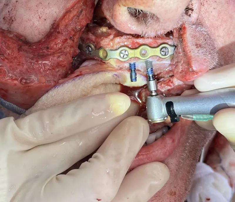
Image content: This image is available to view online.
View image online (https://assets.clevelandclinic.org/transform/422797c9-6b63-4cee-b011-8eb8ec72a4e4/ReconstructionDeficientMaxilla-Tso-CQD-Inset2_800x689)
Dental implants placed during the same operation as the cancer removal and free flap reconstruction
The patient was taken to the operating room a couple of weeks later. He underwent composite resection that included total palatectomy and total septectomy, performed by Eric Lamarre, MD, a staff head and neck surgeon; fibula free flap transfer, performed by Dr. Liu; and dental implant placement, performed by Dr. Tso.
Advertisement
“This was a rapidly progressive cancer, and because of that, we didn't have the luxury of time to do any preoperative surgical planning,” says Dr. Liu. “The design of the free flap reconstruction and implant placement had to be done in real-time intraoperatively with collaboration between myself and Dr. Tso. Although this added operative time to an already lengthy case, it was the most reliable way to achieve dental rehab for this patient.”
The patient wanted to complete as much of the reconstruction as possible during the initial operation, while also preserving facial appearance.
Following these procedures, the patient completed adjuvant radiation. He then underwent revision surgery about six months later to reposition the nose, reshape the buccal vestibule and expose the dental implants. His teeth were placed about a year after the initial surgery.
“Chemoradiation typically begins six weeks after surgery and continues for five to seven weeks,” says Dr. Tso. “After that, the mouth needs time to heal, which can delay dental treatment for six to nine additional months before teeth can be placed.”
Dr. Tso says the patient is doing well and continues to undergo regular monitoring.
“Patients with similar resections often live the remainder of their lives without teeth because conventional dentures do not stay in,” explains Dr. Tso. “Placing implants after chemotherapy and radiation would have been much more complicated because of the need to open up the free flap, lower implant success rates, and the risk of osteoradionecrosis. Including dental surgery at the time of cancer resection sets the patient up for a close to pre-cancer quality of life even after extensive treatment. With advanced tumors, coordinated planning can help preserve meaningful reconstructive and functional options.”
Advertisement
Dr. Tso says these examples underscore the value of multidisciplinary care in complex maxillofacial reconstruction.
“Combining expertise from multiple specialties allows for precise, coordinated treatment that may not be possible within a single specialty,” says Dr. Tso. “Through this level of collaboration, we routinely offer a level of care unavailable at most other institutions.
Advertisement
Neurotology and neurosurgery specialists outline collaborative strategies for preserving function and tailoring treatment
How effective management integrates multiple specialties and therapies
Systematic review indicates more can be done for higher-risk patients
Analysis of HNSCC patients shows HPV status to be predictive of higher abundance of oncobacteria within the tumor
Case study illustrates the potential of a dual-subspecialist approach
Evidence-based recommendations for balancing cancer control with quality of life
Study shows no negative impact for individuals with better contralateral ear performance
HNS device offers new solution for those struggling with CPAP
 Search