Locations:

An isolated nontuberculous mycobacterial vocal fold infection
Advertisement
Cleveland Clinic is a non-profit academic medical center. Advertising on our site helps support our mission. We do not endorse non-Cleveland Clinic products or services. Policy
When laryngitis is the presenting symptom of an infection, delays in diagnosis are common. Here we present the case of a patient who suffered from chronic laryngitis. The 58-year-old patient, who came to us for a second opinion, had a five-year history of vocal fold inflammation and hoarseness. We diagnosed a Mycobacterium Kansasii infection of the laryngopharynx, which we reported recently in Laryngoscope.
As an expatriate healthcare provider, she is widely traveled, and has lived in 20 different countries. It was during her time living in West Africa in 2012 that she first developed laryngitis.
Initial treatment was a course of prednisone, antibiotics and inhalers, and only generated partial improvement, with intermittent bouts of hoarseness. In 2014, a proton pump inhibitor was trialed, but brought no symptom relief. The patient’s symptoms worsened in 2015, at which time she reports experience new symptoms, including low-grade fevers, chills, fatigue, neck pain and night sweats.
Computed tomography scans from 2017 revealed a few nodules on the lungs. Endoscopy from around the same time found some inflammation of the right vocal fold, and a normal left vocal fold. All cultures were negative at this time.
In June 2017, the patient underwent right vocal fold mass excision. Histopathology showed evidence of inflammation, but was otherwise benign.
When the patient sought a second opinion at our facility, she had been hoarse for five years. She spoke with a whisper at all times. Our stroboscopy, performed two months following the excision, showed more discrete inflammation. We tested for tuberculosis and did a complete rheumatologic work-up: both were negative. We prescribed fluconazole and trimethoprim/sulfamethoxazole, but saw no improvement in symptoms.
Advertisement
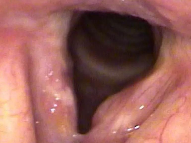
Image content: This image is available to view online.
View image online (https://assets.clevelandclinic.org/transform/b2627af8-b023-46b8-b27e-dca388ed2326/19-ENT-845-benningerLaryng2-Inset_770x579_jpg)
Diffuse vocal fold inflammation with thickening of the right vocal fold. Left vocal fold has a deep submucosal mass with mass effect.
After a repeat stroboscopy, we noted a thickened epithelium on the right vocal fold and a submcosal mass on the left vocal fold. We completed bilateral microflap excision with steroid injection. Biopsy of the vocal fold revealed necrotizing granulomas with rare, acid-fast bacilli upon Ziehl-Neelsen staining.
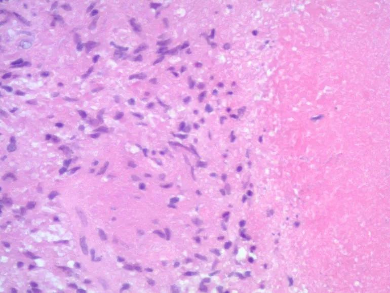
Image content: This image is available to view online.
View image online (https://assets.clevelandclinic.org/transform/06db12d5-4d23-4b6b-ae9f-04f95b12d6cc/19-ENT-845-benningerLaryng1-Inset_770x579_jpg)
Necrotizing granuloma: area of inflammation with necrotizing center.

Image content: This image is available to view online.
View image online (https://assets.clevelandclinic.org/transform/b52ffe0d-a973-4b72-80c8-bbd0361d3209/19-ENT-845-benningerLaryng3-Inset_770x579_jpg)
Ziehl‐Neelsen staining showed rare acid‐fast bacilli with “candy‐cane” striped appearance.
M. Kansasii was identified using broad-range polymerase chain reaction and DNA sequencing. We would not have identified the cause of this infection had we not obtained a tissue culture and stain, and pursued molecular testing.
M. Kansasii generally responds well to treatment, and, once the cause was identified, this case was no different. After six months on a treatment regimen (a combination of isoniazid, rifampin and ethambutol), the patient’s dysphonia improved and most other symptoms resolved.
First isolated in 1953, M. Kansasii is a slow-growing nontuberculous mycobacterial infection that typically presents as a lung infection, with associated cough, chest pain and lung lesions. M. Kansasii infection affecting areas outside the pulmonary system are quite rare. To our knowledge, this is the first reported incidence of an M. Kansasii infection with unique and chronic laryngeal involvement.
Advertisement
Images originally published here:
Lehman B, Procop GW, Silva Merea V, Harrington Sm, Mawhorter SD, Benninger MS. Chronic laryngitis caused by Mycobacterium Kansasii in a traveler. Laryngoscope. 2019 Mar 26. doi: 10.1002/lary.27952. [Epub ahead of print]
Advertisement
Advertisement

How effective management integrates multiple specialties and therapies

Systematic review indicates more can be done for higher-risk patients

Analysis of HNSCC patients shows HPV status to be predictive of higher abundance of oncobacteria within the tumor

Case study illustrates the potential of a dual-subspecialist approach

Evidence-based recommendations for balancing cancer control with quality of life

Study shows no negative impact for individuals with better contralateral ear performance

HNS device offers new solution for those struggling with CPAP

Patient with cerebral palsy undergoes life-saving tumor resection